Structural studies on ADP activation of mammalian glutamate dehydrogenase and the evolution of regulation
Banerjee, S., Schmidt, T., Fang, J., Stanley, C.A., Smith, T.J.(2003) Biochemistry 42: 3446-3456
- PubMed: 12653548
- DOI: https://doi.org/10.1021/bi0206917
- Primary Citation of Related Structures:
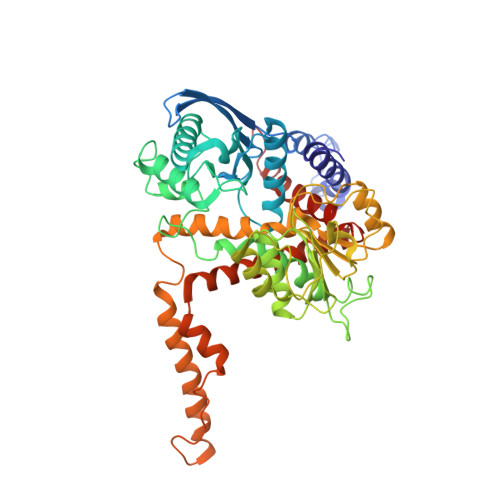
1NQT, 1NR1, 1NR7, 6DHK - PubMed Abstract:
Glutamate dehydrogenase (GDH) is found in all organisms and catalyzes the reversible oxidative deamination of L-glutamate to 2-oxoglutarate. Unlike GDH from bacteria, mammalian GDH exhibits negative cooperativity with respect to coenzyme, activation by ADP, and inhibition by GTP. Presented here are the structures of apo bovine GDH, bovine GDH complexed with ADP, and the R463A mutant form of human GDH (huGDH) that is insensitive to ADP activation. In the absence of active site ligands, the catalytic cleft is in the open conformation, and the hexamers form long polymers in the crystal cell with more interactions than found in the abortive complex crystals. This is consistent with the fact that ADP promotes aggregation in solution. ADP is shown to bind to the second, inhibitory, NADH site yet causes activation. The beta-phosphates of the bound ADP interact with R459 (R463 in huGDH) on the pivot helix. The structure of the ADP-resistant, R463A mutant of human GDH is identical to native GDH with the exception of the truncated side chain on the pivot helix. Together, these results strongly suggest that ADP activates by facilitating the opening of the catalytic cleft. From alignment of GDH from various sources, it is likely that the antenna evolved in the protista prior to the formation of purine regulatory sites. This suggests that there was some selective advantage of the antenna itself and that animals evolved new functions for GDH through the addition of allosteric regulation.
Organizational Affiliation:
Donald Danforth Plant Science Center, St. Louis, Missouri 63132, USA.