1NLD

FAB FRAGMENT OF A NEUTRALIZING ANTIBODY DIRECTED AGAINST AN EPITOPE OF GP41 FROM HIV-1


Experimental Data Snapshot

  • Method: X-RAY DIFFRACTION
  • Resolution: 2.90 Å
  • R-Value Free: 0.326 
  • R-Value Work: 0.198 
  • R-Value Observed: 0.198 

wwPDB Validation   3D Report Full Report


This is version 1.2 of the entry. See complete history


Literature

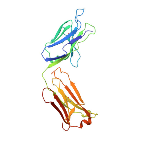
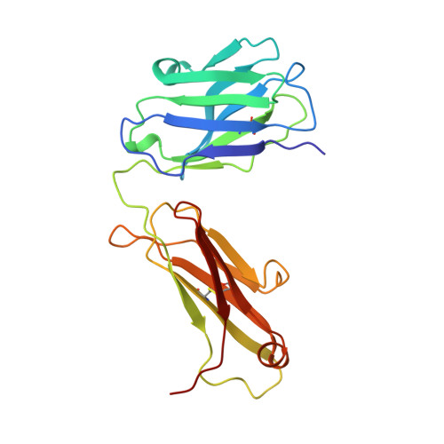
Structure of the Fab fragment from a neutralizing monoclonal antibody directed against an epitope of gp41 from HIV-1.

Davies, C.Beauchamp, J.C.Emery, D.Rawas, A.Muirhead, H.

(1997) Acta Crystallogr D Biol Crystallogr 53: 186-194

  • DOI: https://doi.org/10.1107/S0907444996012048
  • Primary Citation of Related Structures:  
    1NLD

  • PubMed Abstract: 

    The structure of a Fab fragment of a monoclonal antibody (1583) that neutralizes a broad range of HIV-1 isolates has been solved by X-ray crystallography. This antibody is directed against a poliovirus/HIV-I chimaera which presents a conserved epitope of the envelope protein gp41. Crystals of 1583 were obtained in the space group P2(1)2(1)2(1) and the structure solved by molecular replacement. The model has been refined against all data in the range 10-2.9 A to a final crystallographic R factor of 0.198. The antigen-binding site features a well defined groove, typical of antibodies that bind to small antigens, created in part by a relatively short CDR H3. The variable regions of 1583 were sequenced and, given the hydrophilic nature of the epitope, revealed a surprising lack of charged residues in the CDR's. However, the antigen-binding cleft is indeed very polar, due in part to the presence of two charged residues that emanate from outside the recognized CDR's.


  • Organizational Affiliation

    Department of Biochemistry and Molecular Recognition Centre, University of Bristol, England.


Macromolecules
Find similar proteins by:  (by identity cutoff)  |  3D Structure
Entity ID: 1
MoleculeChains Sequence LengthOrganismDetailsImage
FAB1583A [auth L]219Mus musculusMutation(s): 0 
Entity Groups  
Sequence Clusters30% Identity50% Identity70% Identity90% Identity95% Identity100% Identity
Sequence Annotations
Expand
  • Reference Sequence
Find similar proteins by:  (by identity cutoff)  |  3D Structure
Entity ID: 2
MoleculeChains Sequence LengthOrganismDetailsImage
FAB1583B [auth H]215Mus musculusMutation(s): 0 
Entity Groups  
Sequence Clusters30% Identity50% Identity70% Identity90% Identity95% Identity100% Identity
Sequence Annotations
Expand
  • Reference Sequence
Experimental Data & Validation

Experimental Data

  • Method: X-RAY DIFFRACTION
  • Resolution: 2.90 Å
  • R-Value Free: 0.326 
  • R-Value Work: 0.198 
  • R-Value Observed: 0.198 
  • Space Group: P 21 21 21
Unit Cell:
Length ( Å )Angle ( ˚ )
a = 38.15α = 90
b = 82.55β = 90
c = 131γ = 90
Software Package:
Software NamePurpose
XENGENdata collection
X-PLORmodel building
X-PLORrefinement
XENGENdata reduction
X-PLORphasing

Structure Validation

View Full Validation Report


Entry History 

Deposition Data

Revision History  (Full details and data files)

  • Version 1.0: 1996-12-23
    Type: Initial release
  • Version 1.1: 2008-03-24
    Changes: Version format compliance
  • Version 1.2: 2011-07-13
    Changes: Version format compliance