1NJF

Nucleotide bound form of an isolated E. coli clamp loader gamma subunit


Experimental Data Snapshot

  • Method: X-RAY DIFFRACTION
  • Resolution: 2.30 Å
  • R-Value Free: 0.248 
  • R-Value Work: 0.210 
  • R-Value Observed: 0.210 

wwPDB Validation   3D Report Full Report


Ligand Structure Quality Assessment 


This is version 1.4 of the entry. See complete history


Literature

Nucleotide-Induced Conformational Changes in an Isolated Escherichia coli DNA Polymerase III Clamp Loader Subunit

Podobnik, M.Weitze, T.F.O'Donnell, M.Kuriyan, J.

(2003) Structure 11: 253-263

  • DOI: https://doi.org/10.1016/s0969-2126(03)00027-3
  • Primary Citation of Related Structures:  
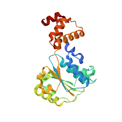
    1NJF, 1NJG

  • PubMed Abstract: 

    Sliding clamps are loaded onto DNA by ATP-driven clamp loader complexes. The structure of the E. coli clamp loader in a nucleotide-free state has been determined previously. We now report crystal structures of a truncated form of the isolated gamma-ATPase subunit, gamma(1-243), of the E. coli clamp loader, in nucleotide-free and bound forms. The gamma subunit adopts a defined conformation when empty, in which the nucleotide binding site is blocked. The binding of either ATPgammaS or ADP, which are shown to bind with equal affinity to gamma(1-243), induces a change in the relative orientation of the two domains such that nucleotides can be accommodated. This change would break one of the gamma:gamma interfaces seen in the empty clamp loader complex, and may represent one step in the activation process.


  • Organizational Affiliation

    Department of Molecular and Cell Biology, Howard Hughes Medical Institute, University of California, Berkeley, Berkeley, CA 94720, USA.


Macromolecules
Find similar proteins by:  (by identity cutoff)  |  3D Structure
Entity ID: 1
MoleculeChains Sequence LengthOrganismDetailsImage
DNA polymerase III subunit gamma
A, B, C, D
250Escherichia coliMutation(s): 0 
Gene Names: DnaX
EC: 2.7.7.7
UniProt
Find proteins for P06710 (Escherichia coli (strain K12))
Explore P06710 
Go to UniProtKB:  P06710
Entity Groups  
Sequence Clusters30% Identity50% Identity70% Identity90% Identity95% Identity100% Identity
UniProt GroupP06710
Sequence Annotations
Expand
  • Reference Sequence
Small Molecules
Ligands 3 Unique
IDChains Name / Formula / InChI Key2D Diagram3D Interactions
AGS
Query on AGS

Download Ideal Coordinates CCD File 
F [auth A],
H [auth B],
L [auth D]
PHOSPHOTHIOPHOSPHORIC ACID-ADENYLATE ESTER
C10 H16 N5 O12 P3 S
NLTUCYMLOPLUHL-KQYNXXCUSA-N
ADP
Query on ADP

Download Ideal Coordinates CCD File 
J [auth C]ADENOSINE-5'-DIPHOSPHATE
C10 H15 N5 O10 P2
XTWYTFMLZFPYCI-KQYNXXCUSA-N
ZN
Query on ZN

Download Ideal Coordinates CCD File 
E [auth A],
G [auth B],
I [auth C],
K [auth D]
ZINC ION
Zn
PTFCDOFLOPIGGS-UHFFFAOYSA-N
Binding Affinity Annotations 
IDSourceBinding Affinity
ADP PDBBind:  1NJF Kd: 1030 (nM) from 1 assay(s)
Binding MOAD:  1NJF Kd: 1030 (nM) from 1 assay(s)
AGS Binding MOAD:  1NJF Kd: 620 (nM) from 1 assay(s)
Experimental Data & Validation

Experimental Data

  • Method: X-RAY DIFFRACTION
  • Resolution: 2.30 Å
  • R-Value Free: 0.248 
  • R-Value Work: 0.210 
  • R-Value Observed: 0.210 
  • Space Group: P 21 21 21
Unit Cell:
Length ( Å )Angle ( ˚ )
a = 71.708α = 90
b = 113.347β = 90
c = 121.355γ = 90
Software Package:
Software NamePurpose
DENZOdata reduction
SCALEPACKdata scaling
MLPHAREphasing
CNSrefinement

Structure Validation

View Full Validation Report


Ligand Structure Quality Assessment 


Entry History 

Deposition Data

Revision History  (Full details and data files)

  • Version 1.0: 2003-04-08
    Type: Initial release
  • Version 1.1: 2008-04-29
    Changes: Version format compliance
  • Version 1.2: 2011-07-13
    Changes: Version format compliance
  • Version 1.3: 2012-03-07
    Changes: Non-polymer description
  • Version 1.4: 2024-02-14
    Changes: Data collection, Database references, Derived calculations