Towards an understanding of the poliovirus replication complex: the solution structure of the soluble domain of the poliovirus 3A protein.
Strauss, D.M., Glustrom, L.W., Wuttke, D.S.(2003) J Mol Biol 330: 225-234
- PubMed: 12823963
- DOI: https://doi.org/10.1016/s0022-2836(03)00577-1
- Primary Citation of Related Structures:
1NG7 - PubMed Abstract:
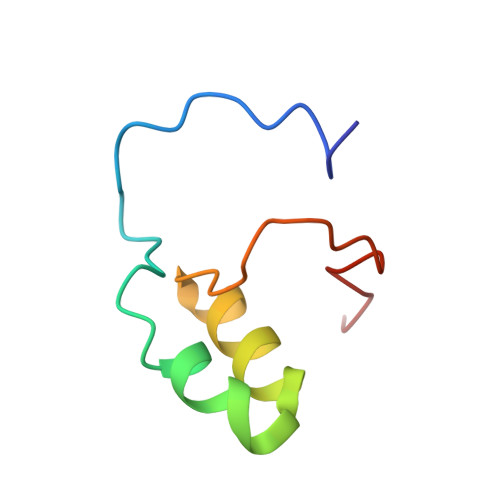
Poliovirus is a positive-strand RNA virus and the prototypical member of the Picornaviridae family. Upon infection, the viral RNA genome is translated from a single open reading frame into a polypeptide which undergoes a series of cleavages to ultimately form four structural and seven non-structural proteins. A replication complex is then formed which replicates the viral genome into negative and positive strands for further translation, replication, and packaging into viral progeny. Poliovirus 3A protein (3A) is a critical component of the viral replication complex and is the putative target of enviroxime, an antiviral drug shown to block viral replication. 3A also inhibits host cell endoplasmic reticulum-to-Golgi apparatus transport, a function which may play a key role in viral evasion from the host immune response. 3A, an 87-residue protein consisting of a soluble N terminus and a hydrophobic C terminus, is formed by the cleavage of the precursor protein 3AB into 3A and 3B (VPg). Although they differ by only 22 residues, the precursor protein 3AB and its cleavage product 3A have distinct functions in viral replication. We have determined the structure of the soluble, N-terminal domain of 3A (3A-N) using NMR spectroscopy. We show that 3A-N exists as a symmetric dimer, and each monomer consists of an alpha-helical hairpin with unstructured, yet functional, N- and C termini. We also show that the 3A-N structure contains a negatively charged surface patch and provides a context for interpreting the biochemical characteristics of a number of previously reported 3A and 3AB mutants.
Organizational Affiliation:
Department of Chemistry and Biochemistry, UCB 215, University of Colorado at Boulder, Boulder, CO 80309-0215, USA.