1NFD

AN ALPHA-BETA T CELL RECEPTOR (TCR) HETERODIMER IN COMPLEX WITH AN ANTI-TCR FAB FRAGMENT DERIVED FROM A MITOGENIC ANTIBODY


Experimental Data Snapshot

  • Method: X-RAY DIFFRACTION
  • Resolution: 2.80 Å
  • R-Value Free: 0.309 
  • R-Value Work: 0.243 
  • R-Value Observed: 0.243 

wwPDB Validation   3D Report Full Report


This is version 2.1 of the entry. See complete history


Literature

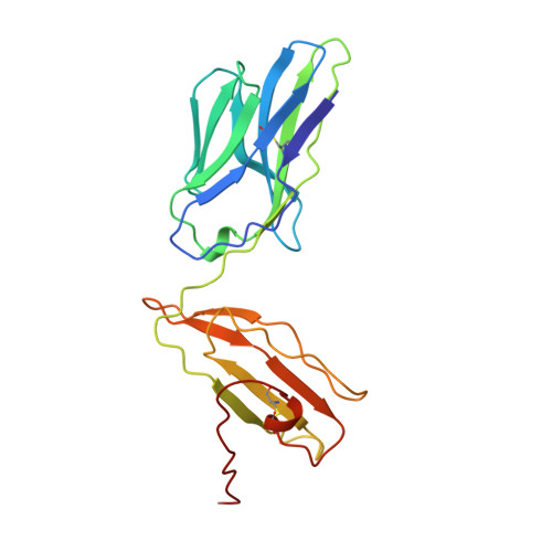
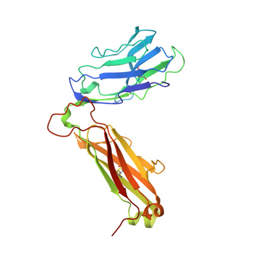
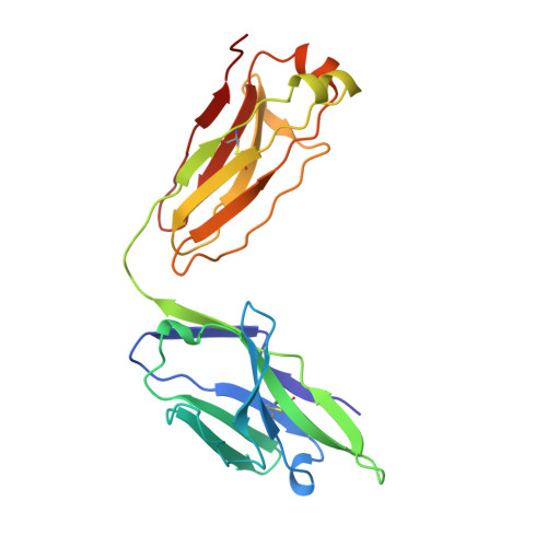
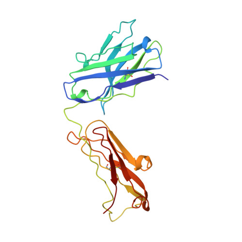
Atomic structure of an alphabeta T cell receptor (TCR) heterodimer in complex with an anti-TCR fab fragment derived from a mitogenic antibody.

Wang, J.Lim, K.Smolyar, A.Teng, M.Liu, J.Tse, A.G.Liu, J.Hussey, R.E.Chishti, Y.Thomson, C.T.Sweet, R.M.Nathenson, S.G.Chang, H.C.Sacchettini, J.C.Reinherz, E.L.

(1998) EMBO J 17: 10-26

  • DOI: https://doi.org/10.1093/emboj/17.1.10
  • Primary Citation of Related Structures:  
    1NFD

  • PubMed Abstract: 

    Each T cell receptor (TCR) recognizes a peptide antigen bound to a major histocompatibility complex (MHC) molecule via a clonotypic alphabeta heterodimeric structure (Ti) non-covalently associated with the monomorphic CD3 signaling components. A crystal structure of an alphabeta TCR-anti-TCR Fab complex shows an Fab fragment derived from the H57 monoclonal antibody (mAb), interacting with the elongated FG loop of the Cbeta domain, situated beneath the Vbeta domain. This loop, along with the partially exposed ABED beta sheet of Cbeta, and glycans attached to both Cbeta and Calpha domains, forms a cavity of sufficient size to accommodate a single non-glycosylated Ig domain such as the CD3epsilon ectodomain. That this asymmetrically localized site is embedded within the rigid constant domain module has implications for the mechanism of signal transduction in both TCR and pre-TCR complexes. Furthermore, quaternary structures of TCRs vary significantly even when they bind the same MHC molecule, as manifested by a unique twisting of the V module relative to the C module.


  • Organizational Affiliation

    Laboratory of Immunobiology, Dana-Farber Cancer Institute, Harvard Medical School, Boston, MA 02115, USA.


Macromolecules
Find similar proteins by:  (by identity cutoff)  |  3D Structure
Entity ID: 1
MoleculeChains Sequence LengthOrganismDetailsImage
N15 ALPHA-BETA T-CELL RECEPTOR
A, C
203Mus musculusMutation(s): 0 
Gene Names: N15 T-CELL RECEPTOR
UniProt
Find proteins for P01849 (Mus musculus)
Explore P01849 
Go to UniProtKB:  P01849
Entity Groups  
Sequence Clusters30% Identity50% Identity70% Identity90% Identity95% Identity100% Identity
UniProt GroupP01849
Sequence Annotations
Expand
  • Reference Sequence
Find similar proteins by:  (by identity cutoff)  |  3D Structure
Entity ID: 2
MoleculeChains Sequence LengthOrganismDetailsImage
N15 ALPHA-BETA T-CELL RECEPTOR
B, D
239Mus musculusMutation(s): 0 
Gene Names: N15 T-CELL RECEPTOR
UniProt
Find proteins for P01852 (Mus musculus)
Explore P01852 
Go to UniProtKB:  P01852
Entity Groups  
Sequence Clusters30% Identity50% Identity70% Identity90% Identity95% Identity100% Identity
UniProt GroupP01852
Sequence Annotations
Expand
  • Reference Sequence
Find similar proteins by:  (by identity cutoff)  |  3D Structure
Entity ID: 3
MoleculeChains Sequence LengthOrganismDetailsImage
H57 FAB
E, G
212Mus musculusMutation(s): 0 
Gene Names: N15 T-CELL RECEPTOR
Entity Groups  
Sequence Clusters30% Identity50% Identity70% Identity90% Identity95% Identity100% Identity
Sequence Annotations
Expand
  • Reference Sequence
Find similar proteins by:  (by identity cutoff)  |  3D Structure
Entity ID: 4
MoleculeChains Sequence LengthOrganismDetailsImage
H57 FAB
F, H
222Mus musculusMutation(s): 0 
Gene Names: N15 T-CELL RECEPTOR
Entity Groups  
Sequence Clusters30% Identity50% Identity70% Identity90% Identity95% Identity100% Identity
Sequence Annotations
Expand
  • Reference Sequence
Small Molecules
Ligands 1 Unique
IDChains Name / Formula / InChI Key2D Diagram3D Interactions
NAG
Query on NAG

Download Ideal Coordinates CCD File 
I [auth A]
J [auth A]
K [auth A]
L [auth B]
M [auth B]
I [auth A],
J [auth A],
K [auth A],
L [auth B],
M [auth B],
N [auth B],
O [auth B],
P [auth C],
Q [auth C],
R [auth C],
S [auth D],
T [auth D],
U [auth D],
V [auth D]
2-acetamido-2-deoxy-beta-D-glucopyranose
C8 H15 N O6
OVRNDRQMDRJTHS-FMDGEEDCSA-N
Experimental Data & Validation

Experimental Data

  • Method: X-RAY DIFFRACTION
  • Resolution: 2.80 Å
  • R-Value Free: 0.309 
  • R-Value Work: 0.243 
  • R-Value Observed: 0.243 
  • Space Group: P 1 21 1
Unit Cell:
Length ( Å )Angle ( ˚ )
a = 74.74α = 90
b = 122.3β = 107.95
c = 115.84γ = 90
Software Package:
Software NamePurpose
DENZOdata reduction
SCALEPACKdata scaling
AMoREphasing
X-PLORmodel building
X-PLORrefinement
X-PLORphasing

Structure Validation

View Full Validation Report


Entry History 

Deposition Data

Revision History  (Full details and data files)

  • Version 1.0: 1998-01-28
    Type: Initial release
  • Version 1.1: 2008-03-03
    Changes: Version format compliance
  • Version 1.2: 2011-07-13
    Changes: Derived calculations, Version format compliance
  • Version 1.3: 2017-07-05
    Changes: Other
  • Version 2.0: 2020-07-29
    Type: Remediation
    Reason: Carbohydrate remediation
    Changes: Advisory, Atomic model, Data collection, Derived calculations, Non-polymer description, Structure summary
  • Version 2.1: 2023-08-09
    Changes: Database references, Refinement description, Structure summary