


Crystal Structure of Nitroreductase from Enterobacter Cloacae
Hecht, H.J., Bryant, C., Erdmann, H., Pelletier, H., Sawaya, R.To be published.
Experimental Data Snapshot
Entity ID: 1 | |||||
|---|---|---|---|---|---|
| Molecule | Chains | Sequence Length | Organism | Details | Image |
| PROTEIN (NITROREDUCTASE) | 216 | Enterobacter cloacae | Mutation(s): 0 | 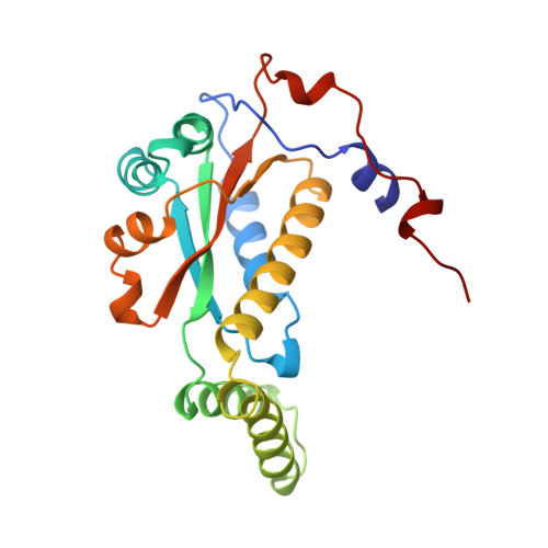 | |
UniProt | |||||
Find proteins for Q01234 (Enterobacter cloacae) Explore Q01234 Go to UniProtKB: Q01234 | |||||
Entity Groups | |||||
| Sequence Clusters | 30% Identity50% Identity70% Identity90% Identity95% Identity100% Identity | ||||
| UniProt Group | Q01234 | ||||
Sequence AnnotationsExpand | |||||
| |||||
| Ligands 1 Unique | |||||
|---|---|---|---|---|---|
| ID | Chains | Name / Formula / InChI Key | 2D Diagram | 3D Interactions | |
| FMN Query on FMN | E [auth A], F [auth B], G [auth C], H [auth D] | FLAVIN MONONUCLEOTIDE C17 H21 N4 O9 P FVTCRASFADXXNN-SCRDCRAPSA-N |  | ||
| Length ( Å ) | Angle ( ˚ ) |
|---|---|
| a = 44 | α = 90 |
| b = 92.5 | β = 93 |
| c = 102.5 | γ = 90 |
| Software Name | Purpose |
|---|---|
| AMoRE | phasing |
| REFMAC | refinement |
| UCSD-system | data reduction |
| UCSD-system | data scaling |
RCSB PDB (citation) is hosted by
RCSB PDB is a member of the

