X-ray structure of a blue-copper nitrite reductase in two crystal forms. The nature of the copper sites, mode of substrate binding and recognition by redox partner.
Dodd, F.E., Van Beeumen, J., Eady, R.R., Hasnain, S.S.(1998) J Mol Biol 282: 369-382
- PubMed: 9735294
- DOI: https://doi.org/10.1006/jmbi.1998.2007
- Primary Citation of Related Structures:
1NDT - PubMed Abstract:
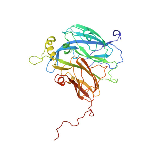
Denitrification is one of the main steps of the global nitrogen cycle that is sustained by prokaryotic organisms. Denitrifying bacteria use two entirely different enzymes in this process, one based on haem cd1 prosthetic groups and the other on type 1-type 2 Cu centres. Copper-containing nitrite reductases (NiRs) are sub-divided into blue and green NiRs, which are respectively thought to be redox partners of azurins and pseudo-azurins. Crystallographic structures of the blue nitrite reductase from Alcaligenes xylosoxidans (AxNiR) are presented in the oxidised hexagonal form and the substrate-bound orthorhombic form to 2.1 A and 2.8 A resolution, respectively. The complete amino acid sequence of AxNiR has been determined by conventional chemical analysis. A 3 A structure of AxNiR has been published where the modelling was based on the sequence of another blue NiR. The higher resolution of the hexagonal form together with the correct sequence allows a detailed comparison with the crystallographic structures of the green NiRs. There is a striking difference in the overall surface charge distribution between the two sub-groups, providing a neat structural explanation for their different reactivities to pseudoazurin or azurin and supporting the view that electron transfer proceeds via complex formation. A detailed examination of the type-1 Cu site, the site responsible for the colour, reveals several subtle differences, including a lateral displacement of 0.7 A for Smet. The structure of the type-2 Cu site, and changes that occur upon substrate binding are discussed in terms of the catalytic mechanism. The similarity of the type 2 Cu site to the catalytic Zn site in carbonic anhydrase and the catalytic Cu site of superoxide dismutase is re-examined in view of the high-resolution (2.1 A) structure.
Organizational Affiliation:
Synchrotron Radiation Department, CCLRC Daresbury Laboratory, Warrington, WA4 4AD, UK.