1NAM

MURINE ALLOREACTIVE SCFV TCR-PEPTIDE-MHC CLASS I MOLECULE COMPLEX


Experimental Data Snapshot

  • Method: X-RAY DIFFRACTION
  • Resolution: 2.70 Å
  • R-Value Free: 0.298 
  • R-Value Work: 0.230 
  • R-Value Observed: 0.237 

wwPDB Validation   3D Report Full Report


This is version 2.1 of the entry. See complete history


Literature

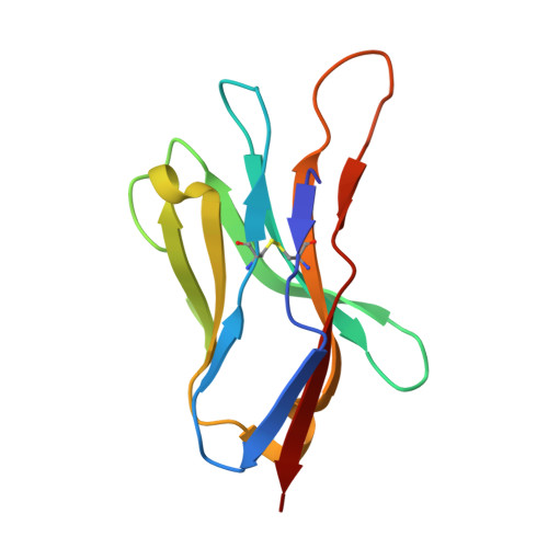
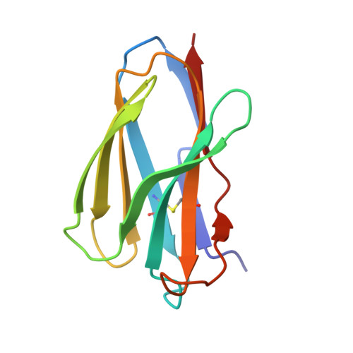
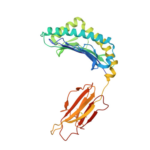
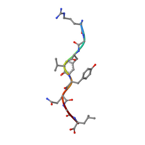
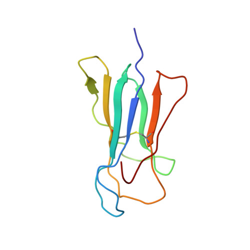
CDR3 loop flexibility contributes to the degeneracy of TCR recognition

Reiser, J.-B.Darnault, C.Gregoire, C.Mosser, T.Mazza, G.Kearnay, A.van der Merwe, P.A.Fontecilla-Camps, J.C.Housset, D.Malissen, B.

(2003) Nat Immunol 4: 241-247

  • DOI: https://doi.org/10.1038/ni891
  • Primary Citation of Related Structures:  
    1NAM, 1NAN

  • PubMed Abstract: 

    T cell receptor (TCR) binding degeneracy lies at the heart of several physiological and pathological phenomena, yet its structural basis is poorly understood. We determined the crystal structure of a complex involving the BM3.3 TCR and an octapeptide (VSV8) bound to the H-2K(b) major histocompatibility complex molecule at a 2.7 A resolution, and compared it with the BM3.3 TCR bound to the H-2K(b) molecule loaded with a peptide that has no primary sequence identity with VSV8. Comparison of these structures showed that the BM3.3 TCR complementarity-determining region (CDR) 3alpha could undergo rearrangements to adapt to structurally different peptide residues. Therefore, CDR3 loop flexibility helps explain TCR binding cross-reactivity.


  • Organizational Affiliation

    Laboratoire de Cristallographie et Cristallogenèse des Protéines, Institut de Biologie Structurale J.-P. Ebel, CEA-CNRS-UJF, 41 rue Jules Horowitz, F-38027 Grenoble Cedex 1, France.


Macromolecules
Find similar proteins by:  (by identity cutoff)  |  3D Structure
Entity ID: 1
MoleculeChains Sequence LengthOrganismDetailsImage
BM3.3 T Cell Receptor alpha-Chain116Mus musculusMutation(s): 0 
UniProt
Find proteins for Q5R1F1 (Mus musculus)
Explore Q5R1F1 
Go to UniProtKB:  Q5R1F1
Entity Groups  
Sequence Clusters30% Identity50% Identity70% Identity90% Identity95% Identity100% Identity
UniProt GroupQ5R1F1
Sequence Annotations
Expand
  • Reference Sequence
Find similar proteins by:  (by identity cutoff)  |  3D Structure
Entity ID: 2
MoleculeChains Sequence LengthOrganismDetailsImage
BM3.3 T Cell Receptor beta-Chain113Mus musculusMutation(s): 0 
UniProt
Find proteins for P04214 (Mus musculus)
Explore P04214 
Go to UniProtKB:  P04214
Entity Groups  
Sequence Clusters30% Identity50% Identity70% Identity90% Identity95% Identity100% Identity
UniProt GroupP04214
Sequence Annotations
Expand
  • Reference Sequence
Find similar proteins by:  (by identity cutoff)  |  3D Structure
Entity ID: 3
MoleculeChains Sequence LengthOrganismDetailsImage
H-2 class I histocompatibility antigen, K-B alpha chain precursorC [auth H]275Mus musculusMutation(s): 0 
Gene Names: H2-K
UniProt
Find proteins for P01901 (Mus musculus)
Explore P01901 
Go to UniProtKB:  P01901
Entity Groups  
Sequence Clusters30% Identity50% Identity70% Identity90% Identity95% Identity100% Identity
UniProt GroupP01901
Sequence Annotations
Expand
  • Reference Sequence

Find similar proteins by:  Sequence   |   3D Structure  

Entity ID: 4
MoleculeChains Sequence LengthOrganismDetailsImage
NucleocapsidD [auth P]8N/AMutation(s): 0 
UniProt
Find proteins for P11212 (Vesicular stomatitis Indiana virus (strain Glasgow))
Explore P11212 
Go to UniProtKB:  P11212
Entity Groups  
UniProt GroupP11212
Sequence Annotations
Expand
  • Reference Sequence
Find similar proteins by:  (by identity cutoff)  |  3D Structure
Entity ID: 5
MoleculeChains Sequence LengthOrganismDetailsImage
Beta-2-microglobulinE [auth L]100Mus musculusMutation(s): 0 
Gene Names: B2M
UniProt & NIH Common Fund Data Resources
Find proteins for P01887 (Mus musculus)
Explore P01887 
Go to UniProtKB:  P01887
IMPC:  MGI:88127
Entity Groups  
Sequence Clusters30% Identity50% Identity70% Identity90% Identity95% Identity100% Identity
UniProt GroupP01887
Sequence Annotations
Expand
  • Reference Sequence
Oligosaccharides

Help

Entity ID: 6
MoleculeChains Length2D Diagram Glycosylation3D Interactions
2-acetamido-2-deoxy-beta-D-glucopyranose-(1-4)-2-acetamido-2-deoxy-beta-D-glucopyranoseF [auth C]2N-Glycosylation
Glycosylation Resources
GlyTouCan:  G42666HT
GlyCosmos:  G42666HT
GlyGen:  G42666HT
Experimental Data & Validation

Experimental Data

  • Method: X-RAY DIFFRACTION
  • Resolution: 2.70 Å
  • R-Value Free: 0.298 
  • R-Value Work: 0.230 
  • R-Value Observed: 0.237 
  • Space Group: P 43 21 2
Unit Cell:
Length ( Å )Angle ( ˚ )
a = 101.86α = 90
b = 101.86β = 90
c = 201.36γ = 90
Software Package:
Software NamePurpose
MOSFLMdata reduction
SCALAdata scaling
AMoREphasing
REFMACrefinement
CCP4data scaling

Structure Validation

View Full Validation Report


Entry History 

Revision History  (Full details and data files)

  • Version 1.0: 2003-03-11
    Type: Initial release
  • Version 1.1: 2008-04-28
    Changes: Version format compliance
  • Version 1.2: 2011-07-13
    Changes: Non-polymer description, Version format compliance
  • Version 2.0: 2020-07-29
    Type: Remediation
    Reason: Carbohydrate remediation
    Changes: Advisory, Atomic model, Data collection, Database references, Derived calculations, Structure summary
  • Version 2.1: 2023-08-16
    Changes: Data collection, Database references, Refinement description, Structure summary