Structure of the functional fragment of auxilin required for catalytic uncoating of clathrin-coated vesicles.
Gruschus, J.M., Han, C.J., Greener, T., Ferretti, J.A., Greene, L.E., Eisenberg, E.(2004) Biochemistry 43: 3111-3119
- PubMed: 15023062
- DOI: https://doi.org/10.1021/bi0354740
- Primary Citation of Related Structures:
1N4C - PubMed Abstract:
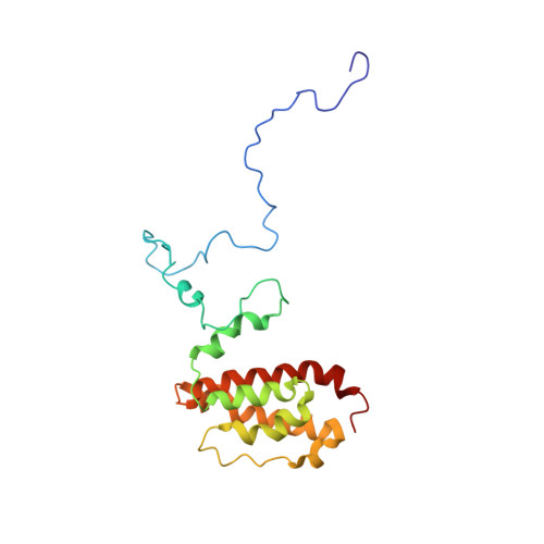
The three-dimensional structure of the C-terminal 20 kDa portion of auxilin, which consists of the clathrin binding region and the C-terminal J-domain, has been determined by NMR. Auxilin is an Hsp40 family protein that catalytically supports the uncoating of clathrin-coated vesicles through recruitment of Hsc70 in an ATP hydrolysis-driven process. This 20 kDa auxilin construct contains the minimal sequential region required to uncoat clathrin-coated vesicles catalytically. The tertiary structure consists of six helices, where the first three are unique to auxilin and believed to be important in the catalytic uncoating of clathrin. The last three helices correspond to the canonical J-domain of Hsp40 proteins. The first helix, helix 1, which contains a conserved FEDLL motif believed to be necessary for clathrin binding, is transient and not packed against the rest of the structure. Helix 1 is joined to helix 2 by a flexible linker. Helix 2 packs loosely against the J-domain surface, whereas helix 3 packs tightly and makes critical contributions to the J-domain core. A long insert loop, also unique to the auxilin J-domain, is seen between helix 4 and helix 5. Comparison with a previously reported structure of auxilin containing only helices 3-6 shows a significant difference in the invariant HPD segment of the J-domain. The region where helix 1 is located corresponds to the expected region of the unstructured G/F-rich domain seen in DnaJ, i.e., the canonical N-terminal J-domain protein. In contrast, the location of helix 1 differs from the substrate binding regions of two other Hsp40 proteins, Escherichia coli Hsc20 and viral large T antigen. The variety of biological functions performed by Hsp40 proteins such as auxilin, as well as the observed differences in the structure and function of their substrate binding regions, supports the notion that Hsp40 proteins act as target-specific adaptors that recruit their more general Hsp70 partners to specific biological roles.
Organizational Affiliation:
Laboratory of Biophysical Chemistry, National Heart, Lung, and Blood Institute, National Institutes of Health, Bethesda, Maryland 20892-0301, USA.