1N3B

Crystal Structure of Dephosphocoenzyme A kinase from Escherichia coli


Experimental Data Snapshot

  • Method: X-RAY DIFFRACTION
  • Resolution: 1.80 Å
  • R-Value Free: 0.246 
  • R-Value Work: 0.217 

wwPDB Validation   3D Report Full Report


This is version 1.4 of the entry. See complete history


Literature

Crystal Structure of a Trimeric Form of Dephosphocoenzyme A Kinase from Escherichia coli

O'Toole, N.Barbosa, J.A.R.G.Li, Y.Hung, L.-W.Matte, A.Cygler, M.

(2003) Protein Sci 12: 327-336

  • DOI: https://doi.org/10.1110/ps.0227803
  • Primary Citation of Related Structures:  
    1N3B

  • PubMed Abstract: 

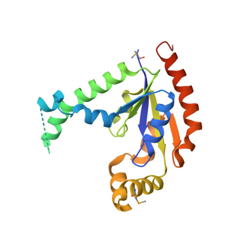
    Coenzyme A (CoA) is an essential cofactor used in a wide variety of biochemical pathways. The final step in the biosynthesis of CoA is catalyzed by dephosphocoenzyme A kinase (DPCK, E.C. 2.7.1.24). Here we report the crystal structure of DPCK from Escherichia coli at 1.8 A resolution. This enzyme forms a tightly packed trimer in its crystal state, in contrast to its observed monomeric structure in solution and to the monomeric, homologous DPCK structure from Haemophilus influenzae. We have confirmed the existence of the trimeric form of the enzyme in solution using gel filtration chromatography measurements. Dephospho-CoA kinase is structurally similar to many nucleoside kinases and other P-loop-containing nucleotide triphosphate hydrolases, despite having negligible sequence similarity to these enzymes. Each monomer consists of five parallel beta-strands flanked by alpha-helices, with an ATP-binding site formed by a P-loop motif. Orthologs of the E. coli DPCK sequence exist in a wide range of organisms, including humans. Multiple alignment of orthologous DPCK sequences reveals a set of highly conserved residues in the vicinity of the nucleotide/CoA binding site.


  • Organizational Affiliation

    Department of Biochemistry, McGill University, Montréal, Québec H3G 1Y6, Canada.


Macromolecules
Find similar proteins by:  (by identity cutoff)  |  3D Structure
Entity ID: 1
MoleculeChains Sequence LengthOrganismDetailsImage
Dephospho-CoA kinase
A, B, C
216Escherichia coliMutation(s): 3 
Gene Names: COAE
EC: 2.7.1.24
UniProt
Find proteins for P0A6I9 (Escherichia coli (strain K12))
Explore P0A6I9 
Go to UniProtKB:  P0A6I9
Entity Groups  
Sequence Clusters30% Identity50% Identity70% Identity90% Identity95% Identity100% Identity
UniProt GroupP0A6I9
Sequence Annotations
Expand
  • Reference Sequence
Small Molecules
Modified Residues  1 Unique
IDChains TypeFormula2D DiagramParent
MSE
Query on MSE
A, B, C
L-PEPTIDE LINKINGC5 H11 N O2 SeMET
Experimental Data & Validation

Experimental Data

  • Method: X-RAY DIFFRACTION
  • Resolution: 1.80 Å
  • R-Value Free: 0.246 
  • R-Value Work: 0.217 
  • Space Group: P 1 21 1
Unit Cell:
Length ( Å )Angle ( ˚ )
a = 55.5α = 90
b = 82.41β = 94.77
c = 76γ = 90
Software Package:
Software NamePurpose
HKL-2000data collection
SCALEPACKdata scaling
SOLVEphasing
CNSrefinement
HKL-2000data reduction

Structure Validation

View Full Validation Report


Entry History 

Revision History  (Full details and data files)

  • Version 1.0: 2003-01-28
    Type: Initial release
  • Version 1.1: 2008-04-28
    Changes: Version format compliance
  • Version 1.2: 2011-07-13
    Changes: Version format compliance
  • Version 1.3: 2014-04-16
    Changes: Data collection
  • Version 1.4: 2017-02-01
    Changes: Structure summary