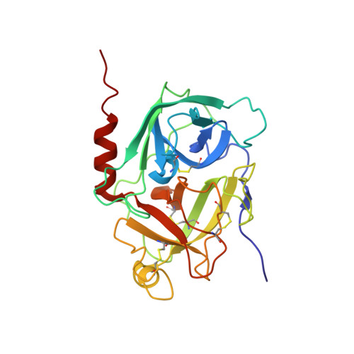
The 2.2-A Crystal Structure of Human Pro-granzyme K Reveals a Rigid Zymogen with Unusual Features
Hink-Schauer, C., Estebanez-Perpina, E., Wilharm, E., Fuentes-Prior, P., Klinkert, W., Bode, W., Jenne, D.E.(2002) J Biol Chem 277: 50923-50933
- PubMed: 12384499
- DOI: https://doi.org/10.1074/jbc.M207962200
- Primary Citation of Related Structures:
1MZA, 1MZD - PubMed Abstract:
Granzyme K (GzmK) belongs to a family of trypsin-like serine proteases localized in electron dense cytoplasmic granules of activated natural killer and cytotoxic T-cells. Like the related granzymes A and B, GzmK can trigger DNA fragmentation and is involved in apoptosis. We expressed the Ser(195) --> Ala variant of human pro-GzmK in Escherichia coli, crystallized it, and determined its 2.2-A x-ray crystal structure. Pro-GzmK possesses a surprisingly rigid structure, which is most similar to activated serine proteases, in particular complement factor D, and not their proforms. The N-terminal peptide Met(14)-Ile(17) projects freely into solution and can be readily approached by cathepsin C, the natural convertase of pro-granzymes. The pre-shaped S1 pocket is occupied by the ion paired residues Lys(188B)-Asp(194) and is hence not available for proper substrate binding. The Ser(214)-Cys(220) segment, which normally provides a template for substrate binding, bulges out of the active site and is distorted. With analogy to complement factor D, we suggest that this strand will maintain its non-productive conformation in mature GzmK, mainly due to the unusual residues Gly(215), Glu(219), and Val(94). We hypothesize that GzmK is proteolytically active only toward specific, as yet unidentified substrates, which upon approach transiently induce a functional active-site conformation.
Organizational Affiliation:
Department of Neuroimmunology, Max-Planck-Institute of Neurobiology, Max-Planck-Institute of Biochemistry, Am Klopferspitz 18a, Planegg-Martinsried D-82152, Germany.