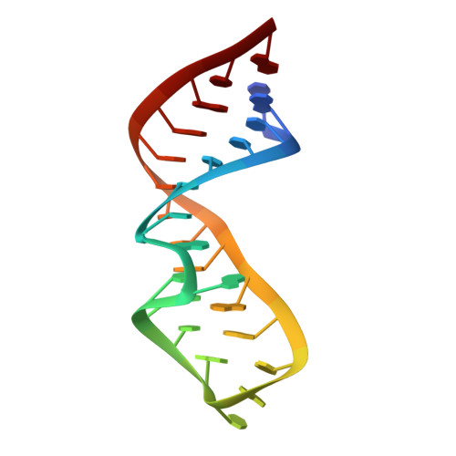
Common and distinctive features of GNRA tetraloops based on a GUAA tetraloop structure at 1.4 A resolution
Correll, C.C., Swinger, K.(2003) RNA 9: 355-363
- PubMed: 12592009
- DOI: https://doi.org/10.1261/rna.2147803
- Primary Citation of Related Structures:
1MSY - PubMed Abstract:
GNRA tetraloops (N is A, C, G, or U; R is A or G) are basic building blocks of RNA structure that often interact with proteins or other RNA structural elements. Understanding sequence-dependent structural variation among different GNRA tetraloops is an important step toward elucidating the molecular basis of specific GNRA tetraloop recognition by proteins and RNAs. Details of the geometry and hydration of this motif have been based on high-resolution crystallographic structures of the GRRA subset of tetraloops; less is known about the GYRA subset (Y is C or U). We report here the structure of a GUAA tetraloop determined to 1.4 A resolution to better define these details and any distinctive features of GYRA tetraloops. The tetraloop is part of a 27-nt structure that mimics the universal sarcin/ricin loop from Escherichia coli 23S ribosomal RNA in which a GUAA tetraloop replaces the conserved GAGA tetraloop. The adenosines of the GUAA tetraloop form an intermolecular contact that is a commonplace RNA tertiary interaction called an A-minor motif. This is the first structure to reveal in great detail the geometry and hydration of a GUAA tetraloop and an A-minor motif. Comparison of tetraloop structures shows a common backbone geometry for each of the eight possible tetraloop sequences and suggests a common hydration. After backbone atom superposition, equivalent bases from different tetraloops unexpectedly depart from coplanarity by as much as 48 degrees. This variation displaces the functional groups of tetraloops implicated in protein and RNA binding, providing a recognition feature.
Organizational Affiliation:
Department of Biochemistry and Molecular Biology, The University of Chicago, Chicago, Illinois 60637, USA. ccorrell@midway.uchicago.edu