1MS9

Triclinic form of Trypanosoma cruzi trans-sialidase, in complex with lactose


Experimental Data Snapshot

  • Method: X-RAY DIFFRACTION
  • Resolution: 1.58 Å
  • R-Value Free: 0.198 
  • R-Value Work: 0.163 
  • R-Value Observed: 0.166 

wwPDB Validation   3D Report Full Report


This is version 2.1 of the entry. See complete history


Literature

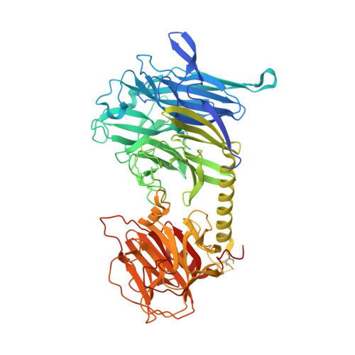
The crystal structure and mode of action of trans-sialidase, a key enzyme of Trypanosoma cruzi pathogenesis

Buschiazzo, A.Amaya, M.F.Cremona, M.L.Frasch, A.C.Alzari, P.M.

(2002) Mol Cell 10: 757-768

  • DOI: https://doi.org/10.1016/s1097-2765(02)00680-9
  • Primary Citation of Related Structures:  
    1MR5, 1MS0, 1MS1, 1MS3, 1MS4, 1MS5, 1MS8, 1MS9

  • PubMed Abstract: 

    Trans-sialidases (TS) are GPI-anchored surface enzymes expressed in specific developmental stages of trypanosome parasites like Trypanosoma cruzi, the etiologic agent of Chagas disease, and T. brucei, the causative agent of sleeping sickness. TS catalyzes the transfer of sialic acid residues from host to parasite glycoconjugates through a transglycosidase reaction that appears to be critical for T. cruzi survival and cell invasion capability. We report here the structure of the T. cruzi trans-sialidase, alone and in complex with sugar ligands. Sialic acid binding is shown to trigger a conformational switch that modulates the affinity for the acceptor substrate and concomitantly creates the conditions for efficient transglycosylation. The structure provides a framework for the structure-based design of novel inhibitors with potential therapeutic applications.


  • Organizational Affiliation

    Unité de Biochimie Structurale, CNRS URA 2185, Institut Pasteur, 25 rue du Dr. Roux, 75724, Paris, France.


Macromolecules
Find similar proteins by:  (by identity cutoff)  |  3D Structure
Entity ID: 1
MoleculeChains Sequence LengthOrganismDetailsImage
trans-sialidase
A, B
648Trypanosoma cruziMutation(s): 7 
EC: 3.2.1.18
UniProt
Find proteins for Q26966 (Trypanosoma cruzi)
Explore Q26966 
Go to UniProtKB:  Q26966
Entity Groups  
Sequence Clusters30% Identity50% Identity70% Identity90% Identity95% Identity100% Identity
UniProt GroupQ26966
Sequence Annotations
Expand
  • Reference Sequence
Oligosaccharides

Help

Entity ID: 2
MoleculeChains Length2D Diagram Glycosylation3D Interactions
beta-D-galactopyranose-(1-4)-beta-D-glucopyranose
C, D
2N/A
Glycosylation Resources
GlyTouCan:  G84224TW
GlyCosmos:  G84224TW
GlyGen:  G84224TW
Biologically Interesting Molecules (External Reference) 1 Unique
Experimental Data & Validation

Experimental Data

  • Method: X-RAY DIFFRACTION
  • Resolution: 1.58 Å
  • R-Value Free: 0.198 
  • R-Value Work: 0.163 
  • R-Value Observed: 0.166 
  • Space Group: P 1
Unit Cell:
Length ( Å )Angle ( ˚ )
a = 51.046α = 86.02
b = 74.204β = 84.17
c = 87.496γ = 88.31
Software Package:
Software NamePurpose
MOSFLMdata reduction
SCALAdata scaling
AMoREphasing
REFMACrefinement
CCP4data scaling

Structure Validation

View Full Validation Report


Entry History 

Deposition Data

Revision History  (Full details and data files)

  • Version 1.0: 2003-03-25
    Type: Initial release
  • Version 1.1: 2008-04-28
    Changes: Version format compliance
  • Version 1.2: 2011-07-13
    Changes: Non-polymer description, Version format compliance
  • Version 2.0: 2020-07-29
    Type: Remediation
    Reason: Carbohydrate remediation
    Changes: Atomic model, Data collection, Database references, Derived calculations, Non-polymer description, Structure summary
  • Version 2.1: 2021-10-27
    Changes: Database references, Structure summary