Solution structure of gamma-bungarotoxin: The functional significance of amino acid residues flanking the RGD motif in integrin binding
Shiu, J.-H., Chen, C.-Y., Chang, L.-S., Chen, Y.-C., Chen, Y.-C., Lo, Y.-H., Liu, Y.-C., Chuang, W.-J.(2004) Proteins 57: 839-849
- PubMed: 15390258
- DOI: https://doi.org/10.1002/prot.20269
- Primary Citation of Related Structures:
1MR6 - PubMed Abstract:
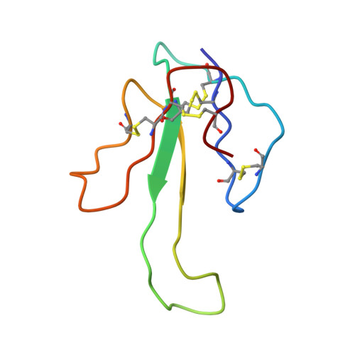
Gamma-bungarotoxin, a snake venom protein isolated from Bungarus multicinctus, contains 68 amino acids, including 10 cysteine residues and a TAVRGDGP sequence at positions 30-37. The solution structure of gamma-bungarotoxin has been determined by nuclear magnetic resonance (NMR) spectroscopy. The structure is similar to that of the short-chain neurotoxins that contain three loops extending from a disulfide-bridged core. The tripeptide Arg-Gly-Asp (RGD) sequence is located at the apex of the flexible loop and is similar to that of other RGD-containing proteins. However, gamma-bungarotoxin only inhibits platelet aggregations with an IC50 of 34 microM. To understand its weak activity in inhibiting platelet aggregation, we mutated the RGD loop sequences of rhodostomin, a potent platelet aggregation inhibitor, from RIPRGDMP to TAVRGDGP, resulting in a 196-fold decrease in activity. In addition, the average Calpha-to-Calpha distance between R33 and G36 of gamma-bungarotoxin is 6.02 A, i.e., shorter than that of other RGD-containing proteins that range from 6.55 to 7.46 A. These results suggested that the amino acid residues flanking the RGD motif might control the width of the RGD loop. This structural difference may be responsible for its decrease in platelet aggregation inhibition compared with other RGD-containing proteins.
Organizational Affiliation:
Department of Biochemistry, National Cheng Kung University College of Medicine, Tainan, Taiwan.