1MQF

Compound I from Proteus mirabilis catalase


Experimental Data Snapshot

  • Method: X-RAY DIFFRACTION
  • Resolution: 2.50 Å
  • R-Value Free: 0.234 
  • R-Value Work: 0.196 
  • R-Value Observed: 0.196 

wwPDB Validation   3D Report Full Report


Ligand Structure Quality Assessment 


This is version 1.2 of the entry. See complete history


Literature

Structural studies of Proteus mirabilis catalase in its ground state, oxidized state and in complex with formic acid.

Andreoletti, P.Pernoud, A.Sainz, G.Gouet, P.Jouve, H.M.

(2003) Acta Crystallogr D Biol Crystallogr 59: 2163-2168

  • DOI: https://doi.org/10.1107/s0907444903019620
  • Primary Citation of Related Structures:  
    1MQF, 1NM0

  • PubMed Abstract: 

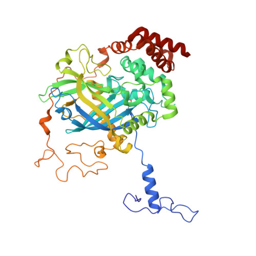
    The structure of Proteus mirabilis catalase in complex with an inhibitor, formic acid, has been solved at 2.3 A resolution. Formic acid is a key ligand of catalase because of its ability to react with the ferric enzyme, giving a high-spin iron complex. Alternatively, it can react with two transient oxidized intermediates of the enzymatic mechanism, compounds I and II. In this work, the structures of native P. mirabilis catalase (PMC) and compound I have also been determined at high resolution (2.0 and 2.5 A, respectively) from frozen crystals. Comparisons between these three PMC structures show that a water molecule present at a distance of 3.5 A from the haem iron in the resting state is absent in the formic acid complex, but reappears in compound I. In addition, movements of solvent molecules are observed during formation of compound I in a cavity located away from the active site, in which a glycerol molecule is replaced by a sulfate. These results give structural insights into the movement of solvent molecules, which may be important in the enzymatic reaction.


  • Organizational Affiliation

    Avidis S.A., Biopôle Clermont-Limagne, 63360 Saint-Beauzire, France.


Macromolecules
Find similar proteins by:  (by identity cutoff)  |  3D Structure
Entity ID: 1
MoleculeChains Sequence LengthOrganismDetailsImage
Catalase484Proteus mirabilisMutation(s): 1 
EC: 1.11.1.6
UniProt
Find proteins for P42321 (Proteus mirabilis)
Explore P42321 
Go to UniProtKB:  P42321
Entity Groups  
Sequence Clusters30% Identity50% Identity70% Identity90% Identity95% Identity100% Identity
UniProt GroupP42321
Sequence Annotations
Expand
  • Reference Sequence
Small Molecules
Ligands 4 Unique
IDChains Name / Formula / InChI Key2D Diagram3D Interactions
HEM
Query on HEM

Download Ideal Coordinates CCD File 
J [auth A]PROTOPORPHYRIN IX CONTAINING FE
C34 H32 Fe N4 O4
KABFMIBPWCXCRK-RGGAHWMASA-L
SO4
Query on SO4

Download Ideal Coordinates CCD File 
B [auth A]
C [auth A]
D [auth A]
E [auth A]
F [auth A]
B [auth A],
C [auth A],
D [auth A],
E [auth A],
F [auth A],
G [auth A],
H [auth A]
SULFATE ION
O4 S
QAOWNCQODCNURD-UHFFFAOYSA-L
GOL
Query on GOL

Download Ideal Coordinates CCD File 
K [auth A]GLYCEROL
C3 H8 O3
PEDCQBHIVMGVHV-UHFFFAOYSA-N
O
Query on O

Download Ideal Coordinates CCD File 
I [auth A]OXYGEN ATOM
O
XLYOFNOQVPJJNP-UHFFFAOYSA-N
Modified Residues  1 Unique
IDChains TypeFormula2D DiagramParent
OMT
Query on OMT
A
L-PEPTIDE LINKINGC5 H11 N O4 SMET
Experimental Data & Validation

Experimental Data

  • Method: X-RAY DIFFRACTION
  • Resolution: 2.50 Å
  • R-Value Free: 0.234 
  • R-Value Work: 0.196 
  • R-Value Observed: 0.196 
  • Space Group: P 62 2 2
Unit Cell:
Length ( Å )Angle ( ˚ )
a = 110α = 90
b = 110β = 90
c = 250γ = 120
Software Package:
Software NamePurpose
MOSFLMdata reduction
SCALAdata scaling
CNSrefinement
CCP4data scaling
CNSphasing

Structure Validation

View Full Validation Report


Ligand Structure Quality Assessment 


Entry History 

Deposition Data

Revision History  (Full details and data files)

  • Version 1.0: 2002-10-02
    Type: Initial release
  • Version 1.1: 2007-10-16
    Changes: Version format compliance
  • Version 1.2: 2011-07-13
    Changes: Derived calculations, Version format compliance