1MNU

UNLIGANDED BACTERICIDAL ANTIBODY AGAINST NEISSERIA MENINGITIDIS


Experimental Data Snapshot

  • Method: X-RAY DIFFRACTION
  • Resolution: 2.50 Å
  • R-Value Free: 0.270 
  • R-Value Work: 0.231 

wwPDB Validation   3D Report Full Report


This is version 1.5 of the entry. See complete history


Literature

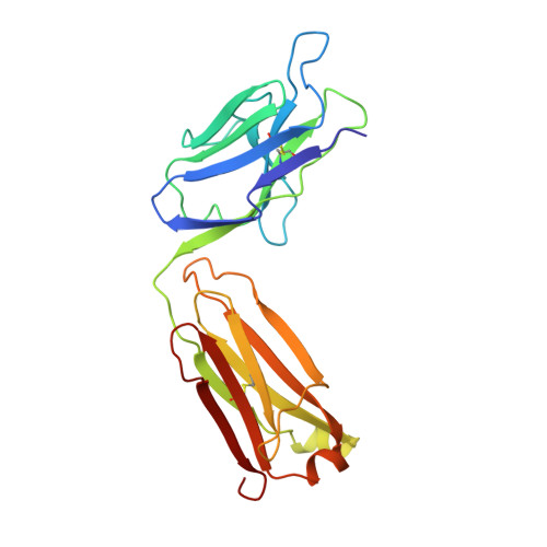
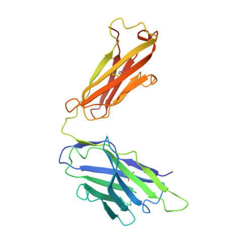
Bactericidal antibody recognition of meningococcal PorA by induced fit. Comparison of liganded and unliganded Fab structures.

van den Elsen, J.Vandeputte-Rutten, L.Kroon, J.Gros, P.

(1999) J Biol Chem 274: 1495-1501

  • DOI: https://doi.org/10.1074/jbc.274.3.1495
  • Primary Citation of Related Structures:  
    1MNU

  • PubMed Abstract: 

    MN12H2 is a bactericidal antibody directed against outer membrane protein PorA epitope P1.16 of Neisseria meningitidis. Binding of MN12H2 to PorA at the meningococcal surface activates the classical complement pathway resulting in bacterial lysis. We have determined the crystal structure of the unliganded MN12H2 Fab fragment in two different crystal forms and compared it with the structure of the Fab in complex with a P1.16-derived peptide. The unliganded Fabs have elbow bend angles of 155 degrees and 159 degrees, whereas the liganded Fab has a more closed elbow bend of 143 degrees. Substantial differences in quaternary and tertiary structure of the antigen binding site are observed between the unliganded and liganded MN12H2 Fab structures that can be attributed to peptide binding. The variable light and heavy chain interface of the liganded Fab is twisted by a 5 degrees rotation along an axis approximately perpendicular to the plane of the interface. Hypervariable loops H1, H2, and framework loop FR-H3 follow this rotation. The hypervariable loop H3 undergoes conformational changes but remains closely linked to hypervariable loop L1. In contrast with the binding site expansion seen in other Fab-peptide structures, the MN12H2 binding site is narrowed upon peptide binding due to the formation of a "false floor" mediated by arginine residue 101 of the light chain. These results indicate that PorA epitope P1.16 of N. meningitidis is recognized by the complement-activating antibody MN12H2 through induced fit, allowing the formation of a highly complementary immune complex.


  • Organizational Affiliation

    Department of Crystal and Structural Chemistry, Bijvoet Center for Biomolecular Research, Utrecht University, Padualaan 8, 3584 CH Utrecht, The Netherlands.


Macromolecules
Find similar proteins by:  (by identity cutoff)  |  3D Structure
Entity ID: 1
MoleculeChains Sequence LengthOrganismDetailsImage
PROTEIN (IGG2A-KAPPA ANTIBODY MN12H2 (LIGHT CHAIN))A [auth L]219Mus musculusMutation(s): 0 
UniProt
Find proteins for A2NHM3 (Mus musculus)
Explore A2NHM3 
Go to UniProtKB:  A2NHM3
Entity Groups  
Sequence Clusters30% Identity50% Identity70% Identity90% Identity95% Identity100% Identity
UniProt GroupA2NHM3
Sequence Annotations
Expand
  • Reference Sequence
Find similar proteins by:  (by identity cutoff)  |  3D Structure
Entity ID: 2
MoleculeChains Sequence LengthOrganismDetailsImage
PROTEIN (IGG2A-KAPPA ANTIBODY MN12H2 (HEAVY CHAIN))B [auth H]225Mus musculusMutation(s): 0 
UniProt
Find proteins for P01863 (Mus musculus)
Explore P01863 
Go to UniProtKB:  P01863
Entity Groups  
Sequence Clusters30% Identity50% Identity70% Identity90% Identity95% Identity100% Identity
UniProt GroupP01863
Sequence Annotations
Expand
  • Reference Sequence
Experimental Data & Validation

Experimental Data

  • Method: X-RAY DIFFRACTION
  • Resolution: 2.50 Å
  • R-Value Free: 0.270 
  • R-Value Work: 0.231 
  • Space Group: C 1 2 1
Unit Cell:
Length ( Å )Angle ( ˚ )
a = 114.617α = 90
b = 85.862β = 122.69
c = 87.133γ = 90
Software Package:
Software NamePurpose
DENZOdata reduction
SCALEPACKdata scaling
CNSrefinement
CNSphasing

Structure Validation

View Full Validation Report


Entry History 

Deposition Data

Revision History  (Full details and data files)

  • Version 1.0: 1999-05-06
    Type: Initial release
  • Version 1.1: 2008-04-26
    Changes: Version format compliance
  • Version 1.2: 2011-07-13
    Changes: Version format compliance
  • Version 1.3: 2018-03-07
    Changes: Data collection
  • Version 1.4: 2019-02-06
    Changes: Data collection, Database references, Experimental preparation, Source and taxonomy
  • Version 1.5: 2023-12-27
    Changes: Data collection, Database references, Derived calculations, Refinement description