1MHP

Crystal structure of a chimeric alpha1 integrin I-domain in complex with the Fab fragment of a humanized neutralizing antibody


Experimental Data Snapshot

  • Method: X-RAY DIFFRACTION
  • Resolution: 2.80 Å
  • R-Value Free: 0.272 
  • R-Value Work: 0.213 
  • R-Value Observed: 0.222 

wwPDB Validation   3D Report Full Report


This is version 1.3 of the entry. See complete history


Literature

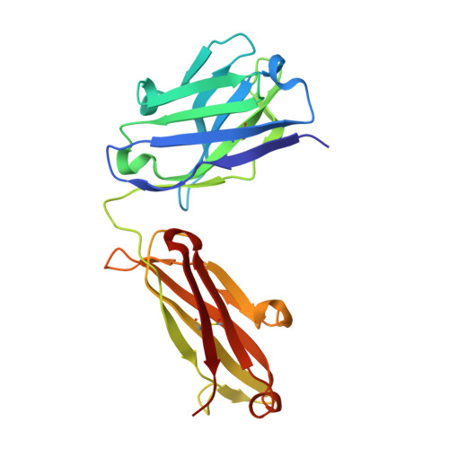
Crystal Structure of the alpha 1 beta 1 Integrin I Domain in Complex with an Antibody Fab Fragment

Karpusas, M.Ferrant, J.Weinreb, P.Carmillo, A.Taylor, F.Garber, E.

(2003) J Mol Biol 327: 1031-1041

  • DOI: https://doi.org/10.1016/s0022-2836(03)00203-1
  • Primary Citation of Related Structures:  
    1MHP

  • PubMed Abstract: 

    The alpha1beta1 (VLA-1) integrin is a cell-surface receptor for collagen and laminin and has been implicated in biological pathways involved in several pathological processes. These processes may be inhibited by the monoclonal antibody AQC2, which binds with high affinity to human alpha1beta1 integrin. To understand the structural basis of the inhibition we determined the crystal structure of the complex of a chimeric rat/human I domain of the alpha1beta1 integrin and the Fab fragment of humanized AQC2 antibody. The structure of the complex shows that the antibody blocks the collagen binding site of the I domain. An aspartate residue, from the CDR3 loop of the antibody heavy chain, coordinates the MIDAS metal ion in a manner similar to that of a glutamate residue from collagen. Substitution of the aspartate residue by alanine or arginine results in significant reduction of antibody binding affinity. Interestingly, although the mode of metal ion coordination resembles that of the open conformation, the I domain maintains an overall closed conformation previously observed only for unliganded I domains.


  • Organizational Affiliation

    Biogen, Inc., 14 Cambridge Center, Cambridge, MA 02142, USA. michael_karpusas@yahoo.com


Macromolecules
Find similar proteins by:  (by identity cutoff)  |  3D Structure
Entity ID: 1
MoleculeChains Sequence LengthOrganismDetailsImage
integrin alpha 1, (RESIDUES 169-360)A,
D [auth B]
192Rattus norvegicusMutation(s): 4 
UniProt
Find proteins for P18614 (Rattus norvegicus)
Explore P18614 
Go to UniProtKB:  P18614
Entity Groups  
Sequence Clusters30% Identity50% Identity70% Identity90% Identity95% Identity100% Identity
UniProt GroupP18614
Sequence Annotations
Expand
  • Reference Sequence
Find similar proteins by:  (by identity cutoff)  |  3D Structure
Entity ID: 2
MoleculeChains Sequence LengthOrganismDetailsImage
Fab fragment, heavy chainB [auth H],
E [auth X]
219Mus musculusMutation(s): 0 
Entity Groups  
Sequence Clusters30% Identity50% Identity70% Identity90% Identity95% Identity100% Identity
Sequence Annotations
Expand
  • Reference Sequence
Find similar proteins by:  (by identity cutoff)  |  3D Structure
Entity ID: 3
MoleculeChains Sequence LengthOrganismDetailsImage
FAB FRAGMENT, light chainC [auth L],
F [auth Y]
212Mus musculusMutation(s): 0 
Entity Groups  
Sequence Clusters30% Identity50% Identity70% Identity90% Identity95% Identity100% Identity
Sequence Annotations
Expand
  • Reference Sequence
Experimental Data & Validation

Experimental Data

  • Method: X-RAY DIFFRACTION
  • Resolution: 2.80 Å
  • R-Value Free: 0.272 
  • R-Value Work: 0.213 
  • R-Value Observed: 0.222 
  • Space Group: P 65
Unit Cell:
Length ( Å )Angle ( ˚ )
a = 255.09α = 90
b = 255.09β = 90
c = 38.64γ = 120
Software Package:
Software NamePurpose
DENZOdata reduction
SCALEPACKdata scaling
AMoREphasing
CNXrefinement

Structure Validation

View Full Validation Report


Entry History 

Deposition Data

Revision History  (Full details and data files)

  • Version 1.0: 2003-04-15
    Type: Initial release
  • Version 1.1: 2008-04-28
    Changes: Version format compliance
  • Version 1.2: 2011-07-13
    Changes: Version format compliance
  • Version 1.3: 2021-10-27
    Changes: Database references, Derived calculations