1MEY

CRYSTAL STRUCTURE OF A DESIGNED ZINC FINGER PROTEIN BOUND TO DNA


Experimental Data Snapshot

  • Method: X-RAY DIFFRACTION
  • Resolution: 2.20 Å
  • R-Value Free: 0.319 
  • R-Value Work: 0.224 
  • R-Value Observed: 0.224 

wwPDB Validation   3D Report Full Report


This is version 1.3 of the entry. See complete history


Literature

A 2.2 A Resolution Crystal Structure of a Designed Zinc Finger Protein Bound to DNA

Kim, C.A.Berg, J.M.

(1996) Nat Struct Biol 3: 940-945

  • DOI: https://doi.org/10.1038/nsb1196-940
  • Primary Citation of Related Structures:  
    1MEY

  • PubMed Abstract: 

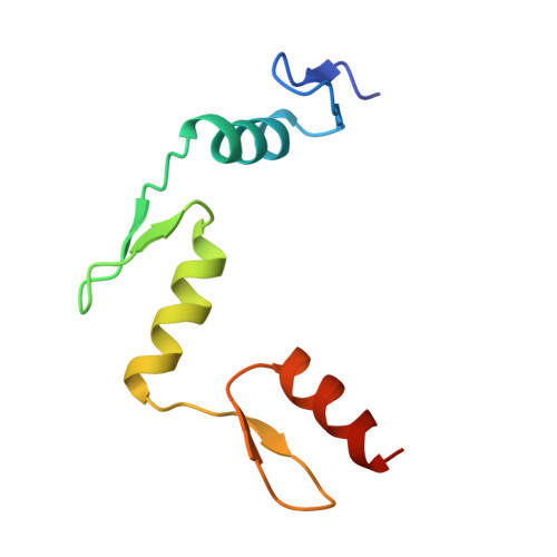
    Considerable recent effort has been devoted to the design and selection of sequence-specific DNA binding proteins based on tandem arrays of Cys2His2 zinc finger domains. While the DNA binding properties of these designed proteins have been studied extensively, the structural basis for site-specific binding has not been examined experimentally. Here we report the crystal structure of a complex between a protein comprised of three consensus-sequence-based zinc finger domains and an oligonucleotide corresponding to a favourable DNA binding site. This structure reveals relatively simple modular interactions and structural adaptations that compensate for differences in contact residue side-chain lengths.


  • Organizational Affiliation

    Department of Chemistry, Johns Hopkins University, Baltimore, Maryland 21218, USA.


Macromolecules

Find similar proteins by:  (by identity cutoff)  |  3D Structure
Entity ID: 3
MoleculeChains Sequence LengthOrganismDetailsImage
CONSENSUS ZINC FINGERE [auth C],
F,
G
87N/AMutation(s): 0 
Entity Groups  
Sequence Clusters30% Identity50% Identity70% Identity90% Identity95% Identity100% Identity
Sequence Annotations
Expand
  • Reference Sequence

Find similar nucleic acids by:  Sequence   |   3D Structure  

Entity ID: 1
MoleculeChains LengthOrganismImage
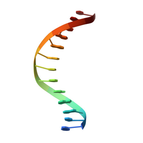
DNA (5'-D(*AP*TP*GP*AP*GP*GP*CP*AP*GP*AP*AP*CP*T)-3')A,
C [auth D]
13N/A
Sequence Annotations
Expand
  • Reference Sequence

Find similar nucleic acids by:  Sequence   |   3D Structure  

Entity ID: 2
MoleculeChains LengthOrganismImage
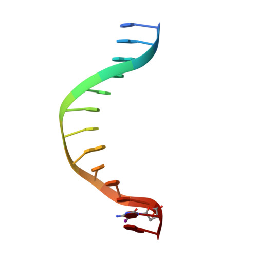
DNA (5'-D(*TP*AP*GP*TP*TP*CP*TP*GP*CP*CP*TP*(C38)P*A)-3')B,
D [auth E]
13N/A
Sequence Annotations
Expand
  • Reference Sequence
Experimental Data & Validation

Experimental Data

  • Method: X-RAY DIFFRACTION
  • Resolution: 2.20 Å
  • R-Value Free: 0.319 
  • R-Value Work: 0.224 
  • R-Value Observed: 0.224 
  • Space Group: P 21 21 21
Unit Cell:
Length ( Å )Angle ( ˚ )
a = 62.07α = 90
b = 165.53β = 90
c = 46.274γ = 90
Software Package:
Software NamePurpose
X-PLORrefinement
PROCESSdata reduction
PROCESSdata scaling

Structure Validation

View Full Validation Report


Entry History 

Deposition Data

Revision History  (Full details and data files)

  • Version 1.0: 1997-03-12
    Type: Initial release
  • Version 1.1: 2008-05-22
    Changes: Version format compliance
  • Version 1.2: 2011-07-13
    Changes: Version format compliance
  • Version 1.3: 2024-02-14
    Changes: Data collection, Database references, Derived calculations