1LSH

LIPID-PROTEIN INTERACTIONS IN LIPOVITELLIN


Experimental Data Snapshot

  • Method: X-RAY DIFFRACTION
  • Resolution: 1.90 Å
  • R-Value Free: 0.255 
  • R-Value Work: 0.193 
  • R-Value Observed: 0.193 

wwPDB Validation   3D Report Full Report


Ligand Structure Quality Assessment 


This is version 2.2 of the entry. See complete history


Literature

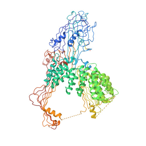
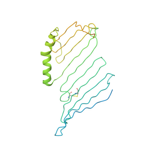
Lipid-protein interactions in lipovitellin.

Thompson, J.R.Banaszak, L.J.

(2002) Biochemistry 41: 9398-9409

  • DOI: https://doi.org/10.1021/bi025674w
  • Primary Citation of Related Structures:  
    1LSH

  • PubMed Abstract: 

    The refined molecular structure of lipovitellin is described using synchrotron cryocrystallographic data to 1.9 A resolution. Lipovitellin is the predominant lipoprotein found in the yolk of egg-laying animals and is involved in lipid and metal storage. It is thought to be related in amino acid sequence to segments of apolipoprotein B and the microsomal transfer protein responsible for the assembly of low-density lipoproteins. Lipovitellin contains a heterogeneous mixture of about 16% (w/w) noncovalently bound lipid, mostly phospholipid. Previous X-ray structural studies at ambient temperature described several different protein domains including a large cavity in each subunit of the dimeric protein. The cavity was free of any visible electron density for lipid molecules at room temperature, suggesting that only dynamic interactions exist with the protein. An important result from this crystallographic study at 100 K is the appearance of some bound ordered lipid along the walls of the binding cavity. The precise identification of the lipid type is difficult because of discontinuities in the electron density. Nonetheless, the conformations of 7 phospholipids and 43 segments of hydrocarbon chains greater than 5 atoms in length have been discovered. The conformations of the bound lipid and the interactions between protein and lipid provide insights into the factors governing lipoprotein formation.


  • Organizational Affiliation

    Department of Biochemistry, Molecular Biology, and Biophysics, University of Minnesota, Minneapolis, Minnesota 55455, USA.


Macromolecules
Find similar proteins by:  (by identity cutoff)  |  3D Structure
Entity ID: 1
MoleculeChains Sequence LengthOrganismDetailsImage
LIPOVITELLIN (LV-1N, LV-1C)1,056Ichthyomyzon unicuspisMutation(s): 0 
Membrane Entity: Yes 
UniProt
Find proteins for Q91062 (Ichthyomyzon unicuspis)
Explore Q91062 
Go to UniProtKB:  Q91062
Entity Groups  
Sequence Clusters30% Identity50% Identity70% Identity90% Identity95% Identity100% Identity
UniProt GroupQ91062
Sequence Annotations
Expand
  • Reference Sequence
Find similar proteins by:  (by identity cutoff)  |  3D Structure
Entity ID: 2
MoleculeChains Sequence LengthOrganismDetailsImage
LIPOVITELLIN (LV-2)319Ichthyomyzon unicuspisMutation(s): 0 
Membrane Entity: Yes 
UniProt
Find proteins for Q91062 (Ichthyomyzon unicuspis)
Explore Q91062 
Go to UniProtKB:  Q91062
Entity Groups  
Sequence Clusters30% Identity50% Identity70% Identity90% Identity95% Identity100% Identity
UniProt GroupQ91062
Sequence Annotations
Expand
  • Reference Sequence
Small Molecules
Ligands 2 Unique
IDChains Name / Formula / InChI Key2D Diagram3D Interactions
PLD
Query on PLD

Download Ideal Coordinates CCD File 
C [auth A]
D [auth A]
E [auth A]
F [auth A]
G [auth A]
C [auth A],
D [auth A],
E [auth A],
F [auth A],
G [auth A],
H [auth A],
MA [auth B]
di-heneicosanoyl phosphatidyl choline
C50 H101 N O8 P
QFFSGJSMHPWZOB-QSCHNALKSA-O
UPL
Query on UPL

Download Ideal Coordinates CCD File 
AA [auth A]
BA [auth A]
CA [auth A]
DA [auth A]
EA [auth A]
AA [auth A],
BA [auth A],
CA [auth A],
DA [auth A],
EA [auth A],
FA [auth A],
GA [auth A],
HA [auth A],
I [auth A],
IA [auth A],
J [auth A],
JA [auth A],
K [auth A],
KA [auth A],
L [auth A],
LA [auth A],
M [auth A],
N [auth A],
NA [auth B],
O [auth A],
OA [auth B],
P [auth A],
PA [auth B],
Q [auth A],
QA [auth B],
R [auth A],
RA [auth B],
S [auth A],
SA [auth B],
T [auth A],
TA [auth B],
U [auth A],
UA [auth B],
V [auth A],
VA [auth B],
W [auth A],
WA [auth B],
X [auth A],
XA [auth B],
Y [auth A],
YA [auth B],
Z [auth A],
ZA [auth B]
UNKNOWN BRANCHED FRAGMENT OF PHOSPHOLIPID
C34 H70
QPCSNFPFMCVVAI-UHFFFAOYSA-N
Modified Residues  1 Unique
IDChains TypeFormula2D DiagramParent
PCA
Query on PCA
A
L-PEPTIDE LINKINGC5 H7 N O3GLN
Experimental Data & Validation

Experimental Data

  • Method: X-RAY DIFFRACTION
  • Resolution: 1.90 Å
  • R-Value Free: 0.255 
  • R-Value Work: 0.193 
  • R-Value Observed: 0.193 
  • Space Group: C 1 2 1
Unit Cell:
Length ( Å )Angle ( ˚ )
a = 190.17α = 90
b = 84.52β = 100.39
c = 89.53γ = 90
Software Package:
Software NamePurpose
HKL-2000data collection
SCALEPACKdata scaling
CNSrefinement
HKL-2000data reduction
CNSphasing

Structure Validation

View Full Validation Report


Ligand Structure Quality Assessment 


Entry History 

Deposition Data

Revision History  (Full details and data files)

  • Version 1.0: 2002-07-31
    Type: Initial release
  • Version 1.1: 2008-04-28
    Changes: Version format compliance
  • Version 1.2: 2011-07-13
    Changes: Version format compliance
  • Version 2.0: 2019-12-25
    Changes: Advisory, Database references, Derived calculations, Polymer sequence, Refinement description
  • Version 2.1: 2021-08-18
    Changes: Advisory, Database references, Derived calculations, Structure summary
  • Version 2.2: 2024-04-03
    Changes: Data collection, Refinement description