1LD9

THE THREE-DIMENSIONAL STRUCTURE OF AN H-2LD PEPTIDE COMPLEX EXPLAINS THE UNIQUE INTERACTION OF LD WITH BETA2M AND PEPTIDE


Experimental Data Snapshot

  • Method: X-RAY DIFFRACTION
  • Resolution: 2.40 Å
  • R-Value Free: 0.279 
  • R-Value Work: 0.186 
  • R-Value Observed: 0.186 

wwPDB Validation   3D Report Full Report


This is version 1.3 of the entry. See complete history


Literature

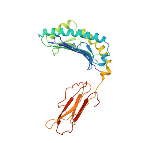
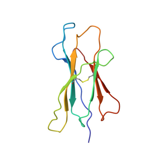
The three-dimensional structure of an H-2Ld-peptide complex explains the unique interaction of Ld with beta-2 microglobulin and peptide.

Balendiran, G.K.Solheim, J.C.Young, A.C.Hansen, T.H.Nathenson, S.G.Sacchettini, J.C.

(1997) Proc Natl Acad Sci U S A 94: 6880-6885

  • DOI: https://doi.org/10.1073/pnas.94.13.6880
  • Primary Citation of Related Structures:  
    1LD9

  • PubMed Abstract: 

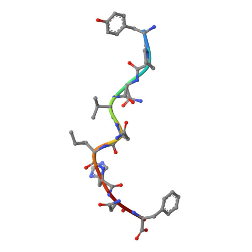
    Solution at 2.5-A resolution of the three-dimensional structure of H-2Ld with a single nine-residue peptide provides a structural basis for understanding its unique interaction with beta-2 microglobulin (beta2m) and peptide. Consistent with the biological data that show an unusually weak association of Ld with beta2m, a novel orientation of the alpha1/alpha2 domains of Ld relative to beta2m results in a dearth of productive contacts compared with other class I proteins. Characteristics of the Ld antigen-binding cleft determine the unique motif of peptides that it binds. Ld has no central anchor residue due to the presence of several bulky side chains in its mid-cleft region. Also, its cleft is significantly more hydrophobic than that of the other class I molecules for which structures are known, resulting in many fewer H-bonds between peptide and cleft residues. The choice of Pro as a consensus anchor at peptide position 2 appears to be related to the hydrophobicity of the B pocket, and to the unique occurrence of Ile (which mirrors Pro in its inability to form H-bonds) at position 63 on the edge of this pocket. Thus, the paucity of stabilizing H-bonds combined with poor complementarity between peptide postion 2 Pro and the B pocket contribute to the weak association between Ld and its peptide antigen. The unique structural interactions of Ld with beta2m and peptide could make Ld more suited than other classical class I molecules to play a role in alternative pathways of antigen presentation.


  • Organizational Affiliation

    Department of Biochemistry and Biophysics, Texas A&M University, College Station, TX 77843, USA.


Macromolecules
Find similar proteins by:  (by identity cutoff)  |  3D Structure
Entity ID: 1
MoleculeChains Sequence LengthOrganismDetailsImage
MHC CLASS I H-2LD HEAVY CHAIN
A, D
268Mus musculusMutation(s): 0 
UniProt
Find proteins for P01897 (Mus musculus)
Explore P01897 
Go to UniProtKB:  P01897
Entity Groups  
Sequence Clusters30% Identity50% Identity70% Identity90% Identity95% Identity100% Identity
UniProt GroupP01897
Sequence Annotations
Expand
  • Reference Sequence
Find similar proteins by:  (by identity cutoff)  |  3D Structure
Entity ID: 2
MoleculeChains Sequence LengthOrganismDetailsImage
BETA-2 MICROGLOBULIN
B, E
99Mus musculusMutation(s): 0 
UniProt & NIH Common Fund Data Resources
Find proteins for P01887 (Mus musculus)
Explore P01887 
Go to UniProtKB:  P01887
IMPC:  MGI:88127
Entity Groups  
Sequence Clusters30% Identity50% Identity70% Identity90% Identity95% Identity100% Identity
UniProt GroupP01887
Sequence Annotations
Expand
  • Reference Sequence

Find similar proteins by:  Sequence   |   3D Structure  

Entity ID: 3
MoleculeChains Sequence LengthOrganismDetailsImage
NANO-PEPTIDE
C, F
9N/AMutation(s): 0 
Sequence Annotations
Expand
  • Reference Sequence
Experimental Data & Validation

Experimental Data

  • Method: X-RAY DIFFRACTION
  • Resolution: 2.40 Å
  • R-Value Free: 0.279 
  • R-Value Work: 0.186 
  • R-Value Observed: 0.186 
  • Space Group: P 21 21 2
Unit Cell:
Length ( Å )Angle ( ˚ )
a = 150.1α = 90
b = 87.2β = 90
c = 80.3γ = 90
Software Package:
Software NamePurpose
XENGENdata collection
XENGENdata reduction
AMoREphasing
X-PLORrefinement
XENGENdata scaling

Structure Validation

View Full Validation Report


Entry History 

Deposition Data

Revision History  (Full details and data files)

  • Version 1.0: 1998-05-06
    Type: Initial release
  • Version 1.1: 2008-03-24
    Changes: Version format compliance
  • Version 1.2: 2011-07-13
    Changes: Version format compliance
  • Version 1.3: 2023-08-09
    Changes: Database references, Refinement description