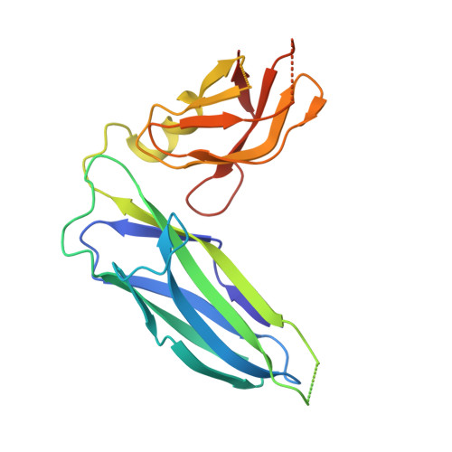
Structure of the S pilus periplasmic chaperone SfaE at 2.2 A resolution.
Knight, S.D., Choudhury, D., Hultgren, S., Pinkner, J., Stojanoff, V., Thompson, A.(2002) Acta Crystallogr D Biol Crystallogr 58: 1016-1022
- PubMed: 12037304
- DOI: https://doi.org/10.1107/s0907444902005954
- Primary Citation of Related Structures:
1L4I - PubMed Abstract:
S pili are sialic acid binding hair-like appendages expressed by pathogenic strains of Escherichia coli. The presence of S pili has been implicated as a virulence factor in both urinary-tract infections and new-born meningitis. Assembly of S pili proceeds via the ubiquitous chaperone/usher pathway. Previously, structures of the homologous chaperones PapD and FimC involved in assembly of P and type-1 pili, respectively, have been solved. Here, the 2.2 A X-ray structure of the S pilus chaperone SfaE is reported. SfaE has the same overall L-shaped structure as PapD and FimC, with two immunoglobulin-like domains oriented at about a 90 degrees angle to each other. Conserved residues in the subunit-binding cleft known to be critical for chaperone function occupy essentially identical positions in SfaE, FimC and PapD. As in free PapD and FimC, the long F1-G1 loop connecting the two last strands of the N-terminal domain is disordered. SfaE crystallizes as a dimer with an extensive dimer interface involving the subunit-binding surfaces of the chaperone. Dimerization via these regions has previously been observed for PapD and might be a general side effect arising from the subunit-binding properties of periplasmic chaperones. The domain interface contains an extended hydrogen-bond network involving three invariant charged residues and two structurally conserved water molecules. It is suggested that disruption of the domain interactions may destabilize the N-terminal domain through exposure of three conserved hydrophobic residues, thereby promoting release of pilus subunits during pilus assembly.
Organizational Affiliation:
Department of Molecular Biology, Uppsala Biomedical Centre, Swedish University of Agricultural Sciences, Box 590, S-753 24 Uppsala, Sweden. stefan@xray.bmc.uu.se