1L3W

C-cadherin Ectodomain


Experimental Data Snapshot

  • Method: X-RAY DIFFRACTION
  • Resolution: 3.08 Å
  • R-Value Free: 0.276 
  • R-Value Work: 0.243 
  • R-Value Observed: 0.243 

wwPDB Validation   3D Report Full Report


Ligand Structure Quality Assessment 


This is version 2.1 of the entry. See complete history


Literature

C-cadherin ectodomain structure and implications for cell adhesion mechanisms

Boggon, T.J.Murray, J.Chappuis-Flament, S.Wong, E.Gumbiner, B.M.Shapiro, L.

(2002) Science 296: 1308-1313

  • DOI: https://doi.org/10.1126/science.1071559
  • Primary Citation of Related Structures:  
    1L3W

  • PubMed Abstract: 

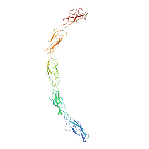
    Cadherins are transmembrane proteins that mediate adhesion between cells in the solid tissues of animals. Here we present the 3.1 angstrom resolution crystal structure of the whole, functional extracellular domain from C-cadherin, a representative "classical" cadherin. The structure suggests a molecular mechanism for adhesion between cells by classical cadherins, and it provides a new framework for understanding both cis (same cell) and trans (juxtaposed cell) cadherin interactions. The trans adhesive interface is a twofold symmetric interaction defined by a conserved tryptophan side chain at the membrane-distal end of a cadherin molecule from one cell, which inserts into a hydrophobic pocket at the membrane-distal end of a cadherin molecule from the opposing cell.


  • Organizational Affiliation

    Department of Biochemistry, Columbia University College of Physicians and Surgeons, 630 West 168th Street, New York, NY 10032, USA.


Macromolecules
Find similar proteins by:  (by identity cutoff)  |  3D Structure
Entity ID: 1
MoleculeChains Sequence LengthOrganismDetailsImage
EP-cadherin546Xenopus laevisMutation(s): 0 
UniProt
Find proteins for P33148 (Xenopus laevis)
Explore P33148 
Go to UniProtKB:  P33148
Entity Groups  
Sequence Clusters30% Identity50% Identity70% Identity90% Identity95% Identity100% Identity
UniProt GroupP33148
Sequence Annotations
Expand
  • Reference Sequence
Small Molecules
Ligands 3 Unique
IDChains Name / Formula / InChI Key2D Diagram3D Interactions
NDG
Query on NDG

Download Ideal Coordinates CCD File 
E [auth A],
L [auth A]
2-acetamido-2-deoxy-alpha-D-glucopyranose
C8 H15 N O6
OVRNDRQMDRJTHS-PVFLNQBWSA-N
NAG
Query on NAG

Download Ideal Coordinates CCD File 
B [auth A]
C [auth A]
D [auth A]
F [auth A]
G [auth A]
B [auth A],
C [auth A],
D [auth A],
F [auth A],
G [auth A],
H [auth A],
I [auth A],
J [auth A],
K [auth A],
M [auth A],
N [auth A],
O [auth A],
P [auth A]
2-acetamido-2-deoxy-beta-D-glucopyranose
C8 H15 N O6
OVRNDRQMDRJTHS-FMDGEEDCSA-N
CA
Query on CA

Download Ideal Coordinates CCD File 
AA [auth A]
BA [auth A]
Q [auth A]
R [auth A]
S [auth A]
AA [auth A],
BA [auth A],
Q [auth A],
R [auth A],
S [auth A],
T [auth A],
U [auth A],
V [auth A],
W [auth A],
X [auth A],
Y [auth A],
Z [auth A]
CALCIUM ION
Ca
BHPQYMZQTOCNFJ-UHFFFAOYSA-N
Experimental Data & Validation

Experimental Data

  • Method: X-RAY DIFFRACTION
  • Resolution: 3.08 Å
  • R-Value Free: 0.276 
  • R-Value Work: 0.243 
  • R-Value Observed: 0.243 
  • Space Group: C 1 2 1
Unit Cell:
Length ( Å )Angle ( ˚ )
a = 127.166α = 90
b = 75.135β = 105.5
c = 129.813γ = 90
Software Package:
Software NamePurpose
DENZOdata reduction
SCALEPACKdata scaling
AMoREphasing
CNSrefinement

Structure Validation

View Full Validation Report


Ligand Structure Quality Assessment 


Entry History 

Deposition Data

Revision History  (Full details and data files)

  • Version 1.0: 2002-04-26
    Type: Initial release
  • Version 1.1: 2008-04-28
    Changes: Version format compliance
  • Version 1.2: 2011-07-13
    Changes: Non-polymer description, Version format compliance
  • Version 1.3: 2011-11-16
    Changes: Atomic model
  • Version 2.0: 2020-07-29
    Type: Remediation
    Reason: Carbohydrate remediation
    Changes: Advisory, Atomic model, Data collection, Database references, Derived calculations, Structure summary
  • Version 2.1: 2023-08-16
    Changes: Data collection, Database references, Refinement description, Structure summary