Crystal structures of mycolic acid cyclopropane synthases from Mycobacterium tuberculosis.
Huang, C.-C., Smith, C.V., Glickman, M.S., Jacobs Jr., W.R., Sacchettini, J.C.(2002) J Biol Chem 277: 11559-11569
- PubMed: 11756461
- DOI: https://doi.org/10.1074/jbc.M111698200
- Primary Citation of Related Structures:
1KP9, 1KPG, 1KPH, 1KPI, 1L1E - PubMed Abstract:
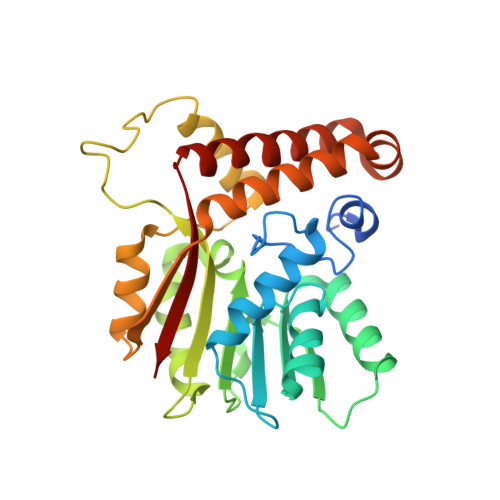
Mycolic acids are major components of the cell wall of Mycobacterium tuberculosis. Several studies indicate that functional groups in the acyl chain of mycolic acids are important for pathogenesis and persistence. There are at least three mycolic acid cyclopropane synthases (PcaA, CmaA1, and CmaA2) that are responsible for these site-specific modifications of mycolic acids. To derive information on the specificity and enzyme mechanism of the family of proteins, the crystal structures of CmaA1, CmaA2, and PcaA were solved to 2-, 2-, and 2.65-A resolution, respectively. All three enzymes have a seven-stranded alpha/beta fold similar to other methyltransferases with the location and interactions with the cofactor S-adenosyl-l-methionine conserved. The structures of the ternary complexes demonstrate the position of the mycolic acid substrate binding site. Close examination of the active site reveals electron density that we believe represents a bicarbonate ion. The structures support the hypothesis that these enzymes catalyze methyl transfer via a carbocation mechanism in which the bicarbonate ion acts as a general base. In addition, comparison of the enzyme structures reveals a possible mechanism for substrate specificity. These structures provide a foundation for rational-drug design, which may lead to the development of new inhibitors effective against persistent bacteria.
Organizational Affiliation:
Department of Biochemistry and Biophysics, Texas A&M University, College Station, Texas 77843-2128, USA.