1KNT

THE 1.6 ANGSTROMS STRUCTURE OF THE KUNITZ-TYPE DOMAIN FROM THE ALPHA3 CHAIN OF THE HUMAN TYPE VI COLLAGEN


Experimental Data Snapshot

  • Method: X-RAY DIFFRACTION
  • Resolution: 1.60 Å
  • R-Value Work: 0.189 
  • R-Value Observed: 0.189 

wwPDB Validation   3D Report Full Report


This is version 1.2 of the entry. See complete history


Literature

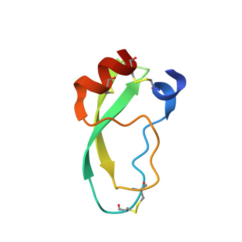
The 1.6 A structure of Kunitz-type domain from the alpha 3 chain of human type VI collagen.

Arnoux, B.Merigeau, K.Saludjian, P.Norris, F.Norris, K.Bjorn, S.Olsen, O.Petersen, L.Ducruix, A.

(1995) J Mol Biol 246: 609-617

  • DOI: https://doi.org/10.1016/s0022-2836(05)80110-x
  • Primary Citation of Related Structures:  
    1KNT

  • PubMed Abstract: 

    The C-terminal Kunitz-type domain from the alpha 3 chain of human type VI collagen (C5), a single 58 amino acid residue chain with three disulfide bridges, was cloned, expressed and crystallized in a monoclonic form, space group P2(1), with a = 25.7 A, b = 38.2 A, c = 28.8 A and beta = 109 degrees. The structure was resolved by molecular replacement, using Alzheimer's protein precursor inhibitor and bovine pancreatic trypsin inhibitor three-dimensional structures as search models. The molecule with one sulfate ion and 43 associated water molecules was refined by XPLOR to an R-factor of 18.9% at 1.6 A. The molecule was not degraded by trypsin and did not inhibit trypsin or tested serine proteases. As opposed to the other Kunitz family members, C5 demonstrates left-handed chirality of the Cys14-Cys38 disulfide bond. Inversion of the Thr13 carbonyl and bulky side-chains at the interface with trypsin in a model of the C5-trypsin complex may explain the lack of inhibition of trypsin.


  • Organizational Affiliation

    Laboratoire de Biologie Structurale, CNRS, Gif sur Yvette, France.


Macromolecules
Find similar proteins by:  (by identity cutoff)  |  3D Structure
Entity ID: 1
MoleculeChains Sequence LengthOrganismDetailsImage
COLLAGEN TYPE VI58Homo sapiensMutation(s): 0 
UniProt & NIH Common Fund Data Resources
Find proteins for P12111 (Homo sapiens)
Explore P12111 
Go to UniProtKB:  P12111
PHAROS:  P12111
GTEx:  ENSG00000163359 
Entity Groups  
Sequence Clusters30% Identity50% Identity70% Identity90% Identity95% Identity100% Identity
UniProt GroupP12111
Sequence Annotations
Expand
  • Reference Sequence
Small Molecules
Ligands 1 Unique
IDChains Name / Formula / InChI Key2D Diagram3D Interactions
SO4
Query on SO4

Download Ideal Coordinates CCD File 
B [auth A]SULFATE ION
O4 S
QAOWNCQODCNURD-UHFFFAOYSA-L
Experimental Data & Validation

Experimental Data

  • Method: X-RAY DIFFRACTION
  • Resolution: 1.60 Å
  • R-Value Work: 0.189 
  • R-Value Observed: 0.189 
  • Space Group: P 1 21 1
Unit Cell:
Length ( Å )Angle ( ˚ )
a = 25.7α = 90
b = 38.2β = 109
c = 28.8γ = 90
Software Package:
Software NamePurpose
X-PLORmodel building
X-PLORrefinement
X-PLORphasing

Structure Validation

View Full Validation Report


Entry History 

Deposition Data

Revision History  (Full details and data files)

  • Version 1.0: 1994-11-01
    Type: Initial release
  • Version 1.1: 2008-03-24
    Changes: Version format compliance
  • Version 1.2: 2011-07-13
    Changes: Version format compliance