1KLT

CRYSTAL STRUCTURE OF PMSF-TREATED HUMAN CHYMASE AT 1.9 ANGSTROMS RESOLUTION


Experimental Data Snapshot

  • Method: X-RAY DIFFRACTION
  • Resolution: 1.90 Å
  • R-Value Free: 0.255 
  • R-Value Work: 0.183 
  • R-Value Observed: 0.183 

wwPDB Validation   3D Report Full Report


This is version 1.4 of the entry. See complete history


Literature

Crystal structure of phenylmethanesulfonyl fluoride-treated human chymase at 1.9 A.

McGrath, M.E.Mirzadegan, T.Schmidt, B.F.

(1997) Biochemistry 36: 14318-14324

  • DOI: https://doi.org/10.1021/bi971403n
  • Primary Citation of Related Structures:  
    1KLT

  • PubMed Abstract: 

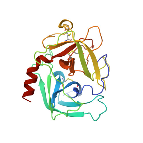
    The X-ray crystal structure of human chymase has been determined to 1.9 A resolution using molecular replacement methods. This first structure of human chymase is present as the Ser 195 ester of alpha-toluenesulfonic acid. The refined structure (Rcryst = 0.183) shows that the inhibitor phenyl moiety lies at the top of the major specificity pocket, S1, while the sulfur is covalently linked to Ser 195-O gamma. The sulfonyl oxygens interact with the oxyanion hole and with His 57-N delta 1. The presence of the inhibitor disturbs the usual gauche position of His 57 and forces it to the trans conformer. Though the primary binding pockets are similarly specific in chymase and chymotrypsin, examination of the extended substrate binding sites reveals the structural basis for chymase's greater discrimination in choosing substrates. The larger 30s loop and its proximity to the active site indicates that it contacts substrate residues C-terminal to the scissile bond. Modeling of substrate at the chymase active site suggests that binding energy may be gained by three main-chain hydrogen bonds provided by substrate residues P2' and P4' and that discriminating interactions with substrate side chains are also likely. The presence of Lys 40 in S1' of human chymase explains its preference for Asp/Glu at P1'. Moreover, the cationic nature of S1' provides a structural basis for human chymase's poor catalytic efficiency when angiotensin II is the substrate.


  • Organizational Affiliation

    Arris Pharmaceutical, South San Francisco, California 94080, USA. mcgrath@arris.com


Macromolecules
Find similar proteins by:  (by identity cutoff)  |  3D Structure
Entity ID: 1
MoleculeChains Sequence LengthOrganismDetailsImage
CHYMASE226Homo sapiensMutation(s): 1 
EC: 3.4.21.39
UniProt & NIH Common Fund Data Resources
Find proteins for P23946 (Homo sapiens)
Explore P23946 
Go to UniProtKB:  P23946
PHAROS:  P23946
GTEx:  ENSG00000092009 
Entity Groups  
Sequence Clusters30% Identity50% Identity70% Identity90% Identity95% Identity100% Identity
UniProt GroupP23946
Sequence Annotations
Expand
  • Reference Sequence
Small Molecules
Ligands 1 Unique
IDChains Name / Formula / InChI Key2D Diagram3D Interactions
PMS
Query on PMS

Download Ideal Coordinates CCD File 
B [auth A]phenylmethanesulfonic acid
C7 H8 O3 S
NIXKBAZVOQAHGC-UHFFFAOYSA-N
Experimental Data & Validation

Experimental Data

  • Method: X-RAY DIFFRACTION
  • Resolution: 1.90 Å
  • R-Value Free: 0.255 
  • R-Value Work: 0.183 
  • R-Value Observed: 0.183 
  • Space Group: P 21 21 21
Unit Cell:
Length ( Å )Angle ( ˚ )
a = 43.93α = 90
b = 58.16β = 90
c = 86.09γ = 90
Software Package:
Software NamePurpose
bioteXdata collection
bioteXdata reduction
X-PLORmodel building
X-PLORrefinement
bioteXdata scaling
X-PLORphasing

Structure Validation

View Full Validation Report


Entry History 

Deposition Data

Revision History  (Full details and data files)

  • Version 1.0: 1998-12-09
    Type: Initial release
  • Version 1.1: 2008-03-24
    Changes: Version format compliance
  • Version 1.2: 2011-07-13
    Changes: Version format compliance
  • Version 1.3: 2021-11-03
    Changes: Database references, Derived calculations, Other, Refinement description
  • Version 1.4: 2024-04-03
    Changes: Data collection, Refinement description