1KJ3

Mhc Class I H-2Kb molecule complexed with pKB1 peptide


Experimental Data Snapshot

  • Method: X-RAY DIFFRACTION
  • Resolution: 2.30 Å
  • R-Value Free: 0.259 
  • R-Value Work: 0.207 
  • R-Value Observed: 0.211 

wwPDB Validation   3D Report Full Report


This is version 1.3 of the entry. See complete history


Literature

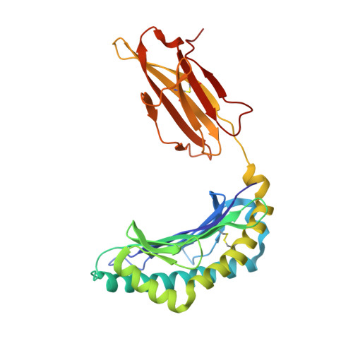
A T cell receptor CDR3beta loop undergoes conformational changes of unprecedented magnitude upon binding to a peptide/MHC class I complex.

Reiser, J.B.Gregoire, C.Darnault, C.Mosser, T.Guimezanes, A.Schmitt-Verhulst, A.M.Fontecilla-Camps, J.C.Mazza, G.Malissen, B.Housset, D.

(2002) Immunity 16: 345-354

  • DOI: https://doi.org/10.1016/s1074-7613(02)00288-1
  • Primary Citation of Related Structures:  
    1KJ3

  • PubMed Abstract: 

    The elongated complementary-determining region (CDR) 3beta found in the unliganded KB5-C20 TCR protrudes from the antigen binding site and prevents its docking onto the peptide/MHC (pMHC) surface according to a canonical diagonal orientation. We now present the crystal structure of a complex involving the KB5-C20 TCR and an octapeptide bound to the allogeneic H-2K(b) MHC class I molecule. This structure reveals how a tremendously large CDR3beta conformational change allows the KB5-C20 TCR to adapt to the rather constrained pMHC surface and achieve a diagonal docking mode. This extreme case of induced fit also shows that TCR plasticity is primarily restricted to CDR3 loops and does not propagate away from the antigen binding site.


  • Organizational Affiliation

    Laboratoire de Cristallographie et Cristallogénèse des Protéines, Institut de Biologie Structurale J.-P. Ebel, CEA-CNRS-UJF, 41 rue Jules Horowitz, F-38027 Grenoble Cedex 1, France.


Macromolecules
Find similar proteins by:  (by identity cutoff)  |  3D Structure
Entity ID: 1
MoleculeChains Sequence LengthOrganismDetailsImage
H-2KB MHC CLASS I MOLECULE ALPHA CHAINA [auth H],
D [auth I]
279Mus musculusMutation(s): 0 
UniProt
Find proteins for P01901 (Mus musculus)
Explore P01901 
Go to UniProtKB:  P01901
Entity Groups  
Sequence Clusters30% Identity50% Identity70% Identity90% Identity95% Identity100% Identity
UniProt GroupP01901
Sequence Annotations
Expand
  • Reference Sequence

Find similar proteins by:  Sequence   |   3D Structure  

Entity ID: 2
MoleculeChains Sequence LengthOrganismDetailsImage
NATURALLY PROCESSED OCTAPEPTIDE PKB1B [auth P],
E [auth Q]
8N/AMutation(s): 0 
UniProt
Find proteins for O08582 (Mus musculus)
Explore O08582 
Go to UniProtKB:  O08582
Entity Groups  
UniProt GroupO08582
Sequence Annotations
Expand
  • Reference Sequence
Find similar proteins by:  (by identity cutoff)  |  3D Structure
Entity ID: 3
MoleculeChains Sequence LengthOrganismDetailsImage
BETA-2 MICROGLOBULINC [auth L],
F [auth M]
99Mus musculusMutation(s): 0 
UniProt & NIH Common Fund Data Resources
Find proteins for P01887 (Mus musculus)
Explore P01887 
Go to UniProtKB:  P01887
IMPC:  MGI:88127
Entity Groups  
Sequence Clusters30% Identity50% Identity70% Identity90% Identity95% Identity100% Identity
UniProt GroupP01887
Sequence Annotations
Expand
  • Reference Sequence
Experimental Data & Validation

Experimental Data

  • Method: X-RAY DIFFRACTION
  • Resolution: 2.30 Å
  • R-Value Free: 0.259 
  • R-Value Work: 0.207 
  • R-Value Observed: 0.211 
  • Space Group: P 1 21 1
Unit Cell:
Length ( Å )Angle ( ˚ )
a = 66.24α = 90
b = 90.63β = 111.51
c = 89.58γ = 90
Software Package:
Software NamePurpose
MOSFLMdata reduction
SCALAdata scaling
AMoREphasing
REFMACrefinement
CCP4data scaling

Structure Validation

View Full Validation Report


Entry History 

Revision History  (Full details and data files)

  • Version 1.0: 2002-03-27
    Type: Initial release
  • Version 1.1: 2008-04-27
    Changes: Version format compliance
  • Version 1.2: 2011-07-13
    Changes: Version format compliance
  • Version 1.3: 2023-08-16
    Changes: Data collection, Database references, Refinement description