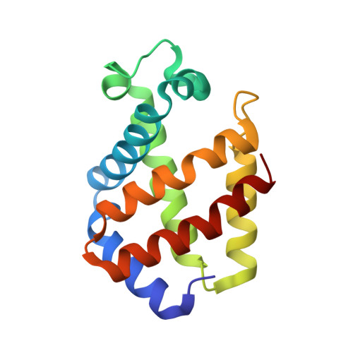
1KFR

Structural plasticity in the eight-helix fold of a trematode hemoglobin


Experimental Data Snapshot

  • Method: X-RAY DIFFRACTION
  • Resolution: 1.85 Å
  • R-Value Free: 0.220 
  • R-Value Work: 0.161 
  • R-Value Observed: 0.164 

wwPDB Validation   3D Report Full Report


Ligand Structure Quality Assessment 


This is version 1.3 of the entry. See complete history


Literature

Structural plasticity in the eight-helix fold of a trematode haemoglobin.

Milani, M.Pesce, A.Dewilde, S.Ascenzi, P.Moens, L.Bolognesi, M.

(2002) Acta Crystallogr D Biol Crystallogr 58: 719-722

  • DOI: https://doi.org/10.1107/s0907444902001865
  • Primary Citation of Related Structures:  
    1KFR

  • PubMed Abstract: 

    The three-dimensional structure of recombinant haemoglobin from the trematode Paramphistomum epiclitum, displaying the highest oxygen affinity so far observed for (non)vertebrate haemoglobins, has previously been determined at 1.17 A resolution (orthorhombic space group P2(1)2(1)2(1)). In the present communication, the three-dimensional structure of wild-type P. epiclitum haemoglobin is reported at 1.85 A resolution in a monoclinic crystal form (R factor = 16.1%, R(free) = 22.0%). Comparison of P. epiclitum (recombinant versus wild-type ferric Hb) structures in the two crystal forms shows structural differences in the haem proximal and distal sites which have not been reported for other known haemoglobin structures previously.


  • Organizational Affiliation

    Department of Physics-INFM, Advanced Biotechnology Centre, University of Genova, Largo Rosanna Benzi 10, I-16146 Genova, Italy.


Macromolecules
Find similar proteins by:  (by identity cutoff)  |  3D Structure
Entity ID: 1
MoleculeChains Sequence LengthOrganismDetailsImage
Hemoglobin147Paramphistomum epiclitumMutation(s): 0 
UniProt
Find proteins for P80721 (Paramphistomum epiclitum)
Explore P80721 
Go to UniProtKB:  P80721
Entity Groups  
Sequence Clusters30% Identity50% Identity70% Identity90% Identity95% Identity100% Identity
UniProt GroupP80721
Sequence Annotations
Expand
  • Reference Sequence
Experimental Data & Validation

Experimental Data

  • Method: X-RAY DIFFRACTION
  • Resolution: 1.85 Å
  • R-Value Free: 0.220 
  • R-Value Work: 0.161 
  • R-Value Observed: 0.164 
  • Space Group: P 1 21 1
Unit Cell:
Length ( Å )Angle ( ˚ )
a = 41.117α = 90
b = 31.45β = 95.5
c = 54.952γ = 90
Software Package:
Software NamePurpose
EPMRphasing
REFMACrefinement
DENZOdata reduction
SCALEPACKdata scaling

Structure Validation

View Full Validation Report


Ligand Structure Quality Assessment 


Entry History 

Deposition Data

Revision History  (Full details and data files)

  • Version 1.0: 2002-04-24
    Type: Initial release
  • Version 1.1: 2008-04-27
    Changes: Version format compliance
  • Version 1.2: 2011-07-13
    Changes: Version format compliance
  • Version 1.3: 2024-02-07
    Changes: Data collection, Database references, Derived calculations