1KDQ

Crystal Structure Analysis of the Mutant S189D Rat Chymotrypsin


Experimental Data Snapshot

  • Method: X-RAY DIFFRACTION
  • Resolution: 2.55 Å
  • R-Value Free: 0.277 
  • R-Value Work: 0.221 
  • R-Value Observed: 0.221 

wwPDB Validation   3D Report Full Report


This is version 1.4 of the entry. See complete history


Literature

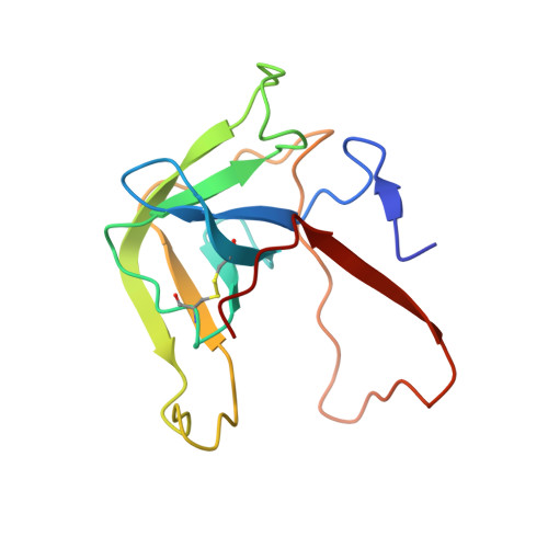
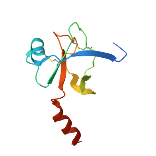
Three Dimensional Structures of S189D Chymotrypsin and D189S Trypsin Mutants: The Effect of Polarity at Site 189 on a Protease-specific Stabilization of the Substrate-binding Site

Szabo, E.Venekei, I.Bocskei, Z.Naray-Szabo, G.Graf, L.

(2003) J Mol Biol 331: 1121-1130

  • DOI: https://doi.org/10.1016/s0022-2836(03)00849-0
  • Primary Citation of Related Structures:  
    1KDQ

  • PubMed Abstract: 

    The crystal structure of S189D rat chymotrypsin have been determined (resolution 2.55A) and compared, together with D189S rat trypsin to wild-type structures to examine why these single mutations resulted in poorly active, non-specific enzymes instead of converting the specificities of trypsin and chymotrypsin into each other. Both mutants have stable structure but suffer from a surprisingly large number of serious deformations. These are restricted to the activation domain, mainly to the substrate-binding region and are larger in S189D chymotrypsin. A wild-type substrate-binding mode in the mutants is disfavored by substantial displacements of the Cys191-Cys220 disulfide and loop segments 185-195 (loop C2/D2) and 217-224 (loop E2/F2) at the specificity site. As a consequence, the substrate-binding clefts become wider and more solvent-accessible in the middle third and occluded in the lower third. Interestingly, while the Ser189 residue in D189S trypsin adopts a chymotrypsin-like conformation, the Asp189 residue in S189D chymotrypsin is turned out toward the solvent. The rearrangements in D189S trypsin are at the same sites where trypsin and trypsinogen differ and, in S189D chymotrypsin, the oxyanion hole as well as the salt-bridge between Asp194 and the N-terminal of Ile16 are missing as in chymotrypsinogen. Despite these similarities, the mutants do not have zymogen conformation. The Ser189Asp and Asp189Ser substitutions are structurally so disruptive probably because the stabilization of such a different specificity site polarities as those after the removal or introduction of a charged residue are beyond the capability of the wild-type conformation of the substrate-binding region.


  • Organizational Affiliation

    Department of Biochemistry, Hungarian Academy of Sciences, Eötvös Loránd University, 1117, Pázmány Péter sétány 1/A, Budapest, Hungary.


Macromolecules
Find similar proteins by:  (by identity cutoff)  |  3D Structure
Entity ID: 1
MoleculeChains Sequence LengthOrganismDetailsImage
CHYMOTRYPSIN B, B CHAIN131Rattus norvegicusMutation(s): 1 
EC: 3.4.21.1
UniProt
Find proteins for P07338 (Rattus norvegicus)
Explore P07338 
Go to UniProtKB:  P07338
Entity Groups  
Sequence Clusters30% Identity50% Identity70% Identity90% Identity95% Identity100% Identity
UniProt GroupP07338
Sequence Annotations
Expand
  • Reference Sequence
Find similar proteins by:  (by identity cutoff)  |  3D Structure
Entity ID: 2
MoleculeChains Sequence LengthOrganismDetailsImage
CHYMOTRYPSIN B, C CHAIN99Rattus norvegicusMutation(s): 1 
EC: 3.4.21.1
UniProt
Find proteins for P07338 (Rattus norvegicus)
Explore P07338 
Go to UniProtKB:  P07338
Entity Groups  
Sequence Clusters30% Identity50% Identity70% Identity90% Identity95% Identity100% Identity
UniProt GroupP07338
Sequence Annotations
Expand
  • Reference Sequence
Small Molecules
Ligands 1 Unique
IDChains Name / Formula / InChI Key2D Diagram3D Interactions
CA
Query on CA

Download Ideal Coordinates CCD File 
C [auth A]CALCIUM ION
Ca
BHPQYMZQTOCNFJ-UHFFFAOYSA-N
Experimental Data & Validation

Experimental Data

  • Method: X-RAY DIFFRACTION
  • Resolution: 2.55 Å
  • R-Value Free: 0.277 
  • R-Value Work: 0.221 
  • R-Value Observed: 0.221 
  • Space Group: C 2 2 21
Unit Cell:
Length ( Å )Angle ( ˚ )
a = 74.589α = 90
b = 76.707β = 90
c = 83.478γ = 90
Software Package:
Software NamePurpose
MOSFLMdata reduction
SCALAdata scaling
AMoREphasing
X-PLORrefinement
CCP4data scaling

Structure Validation

View Full Validation Report


Entry History 

Deposition Data

Revision History  (Full details and data files)

  • Version 1.0: 2003-06-10
    Type: Initial release
  • Version 1.1: 2008-04-27
    Changes: Version format compliance
  • Version 1.2: 2011-07-13
    Changes: Version format compliance
  • Version 1.3: 2018-04-04
    Changes: Data collection
  • Version 1.4: 2021-10-27
    Changes: Database references, Derived calculations