1KCR

CRYSTAL STRUCTURE OF ANTIBODY PC283 IN COMPLEX WITH PS1 PEPTIDE


Experimental Data Snapshot

  • Method: X-RAY DIFFRACTION
  • Resolution: 2.90 Å
  • R-Value Free: 0.277 
  • R-Value Work: 0.184 

wwPDB Validation   3D Report Full Report


This is version 1.2 of the entry. See complete history


Literature

Crystal structure of an antibody bound to an immunodominant peptide epitope: novel features in peptide-antibody recognition.

Nair, D.T.Singh, K.Sahu, N.Rao, K.V.Salunke, D.M.

(2000) J Immunol 165: 6949-6955

  • DOI: https://doi.org/10.4049/jimmunol.165.12.6949
  • Primary Citation of Related Structures:  
    1KCR

  • PubMed Abstract: 

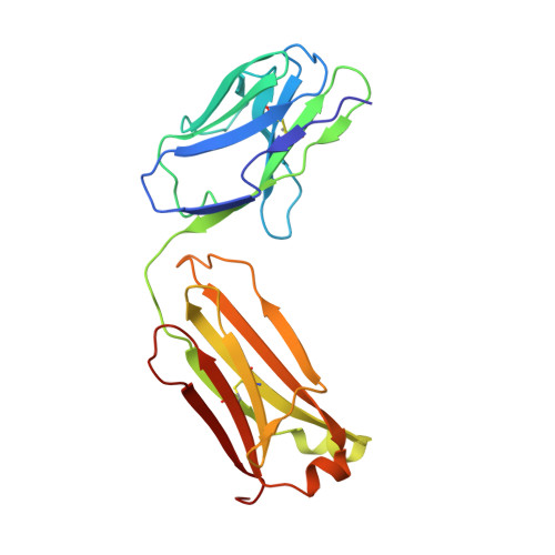
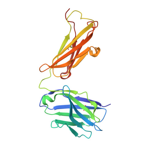
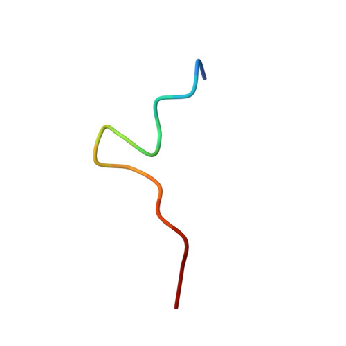
    The crystal structure of Fab of an Ab PC283 complexed with its corresponding peptide Ag, PS1 (HQLDPAFGANSTNPD), derived from the hepatitis B virus surface Ag was determined. The PS1 stretch Gln2P to Phe7P is present in the Ag binding site of the Ab, while the next three residues of the peptide are raised above the binding groove. The residues Ser11P, Thr12P, and Asn13P then loop back onto the Ag-binding site of the Ab. The last two residues, Pro14P and Asp15P, extend outside the binding site without forming any contacts with the Ab. The PC283-PS1 complex is among the few examples where the light chain complementarity-determining regions show more interactions than the heavy chain complementarity-determining regions, and a distal framework residue is involved in Ag binding. As seen from the crystal structure, most of the contacts between peptide and Ab are through the five residues, Leu3-Asp4-Pro5-Ala6-Phe7, of PS1. The paratope is predominantly hydrophobic with aromatic residues lining the binding pocket, although a salt bridge also contributes to stabilizing the Ag-Ab interaction. The molecular surface area buried upon PS1 binding is 756 A(2) for the peptide and 625 A(2) for the Fab, which is higher than what has been seen to date for Ab-peptide complexes. A comparison between PC283 structure and a homology model of its germline ancestor suggests that paratope optimization for PS1 occurs by improving both charge and shape complementarity.


  • Organizational Affiliation

    National Institute of Immunology and International Center of Genetic Engineering and Biotechnology, New Delhi, India.


Macromolecules
Find similar proteins by:  (by identity cutoff)  |  3D Structure
Entity ID: 1
MoleculeChains Sequence LengthOrganismDetailsImage
PC283 IMMUNOGLOBULINA [auth L]213Mus musculusMutation(s): 0 
UniProt
Find proteins for P01837 (Mus musculus)
Explore P01837 
Go to UniProtKB:  P01837
Entity Groups  
Sequence Clusters30% Identity50% Identity70% Identity90% Identity95% Identity100% Identity
UniProt GroupP01837
Sequence Annotations
Expand
  • Reference Sequence
Find similar proteins by:  (by identity cutoff)  |  3D Structure
Entity ID: 2
MoleculeChains Sequence LengthOrganismDetailsImage
PC283 IMMUNOGLOBULINB [auth H]218Mus musculusMutation(s): 0 
UniProt
Find proteins for P01869 (Mus musculus)
Explore P01869 
Go to UniProtKB:  P01869
Entity Groups  
Sequence Clusters30% Identity50% Identity70% Identity90% Identity95% Identity100% Identity
UniProt GroupP01869
Sequence Annotations
Expand
  • Reference Sequence

Find similar proteins by:  Sequence   |   3D Structure  

Entity ID: 3
MoleculeChains Sequence LengthOrganismDetailsImage
PS1 peptideC [auth P]15N/AMutation(s): 0 
UniProt
Find proteins for Q91C35 (Hepatitis B virus genotype A1 subtype adw2 (isolate South Africa/84/2001))
Explore Q91C35 
Go to UniProtKB:  Q91C35
Entity Groups  
Sequence Clusters30% Identity50% Identity70% Identity90% Identity95% Identity100% Identity
UniProt GroupQ91C35
Sequence Annotations
Expand
  • Reference Sequence
Experimental Data & Validation

Experimental Data

  • Method: X-RAY DIFFRACTION
  • Resolution: 2.90 Å
  • R-Value Free: 0.277 
  • R-Value Work: 0.184 
  • Space Group: P 21 21 21
Unit Cell:
Length ( Å )Angle ( ˚ )
a = 69.724α = 90
b = 72.686β = 90
c = 84.493γ = 90
Software Package:
Software NamePurpose
MARdata collection
SCALEPACKdata scaling
AMoREphasing
CNSrefinement
MARdata reduction

Structure Validation

View Full Validation Report


Entry History 

Deposition Data

Revision History  (Full details and data files)

  • Version 1.0: 2002-05-11
    Type: Initial release
  • Version 1.1: 2008-04-27
    Changes: Version format compliance
  • Version 1.2: 2011-07-13
    Changes: Version format compliance