1K40

crystal structure of the FAT domain of focal adhesion kinase


Experimental Data Snapshot

  • Method: X-RAY DIFFRACTION
  • Resolution: 2.25 Å
  • R-Value Free: 0.294 
  • R-Value Work: 0.236 
  • R-Value Observed: 0.236 

wwPDB Validation   3D Report Full Report


This is version 1.3 of the entry. See complete history


Literature

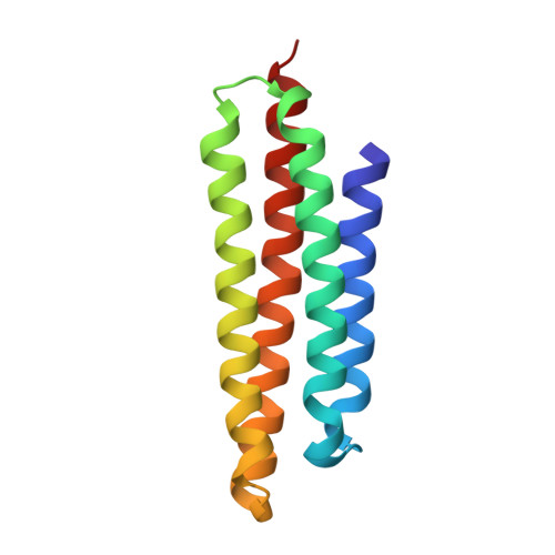
The focal adhesion targeting (FAT) region of focal adhesion kinase is a four-helix bundle that binds paxillin

Hayashi, I.Vuori, K.Liddington, R.C.

(2002) Nat Struct Biol 9: 101-106

  • DOI: https://doi.org/10.1038/nsb755
  • Primary Citation of Related Structures:  
    1K40

  • PubMed Abstract: 

    Focal adhesion kinase (FAK) is a tyrosine kinase found in focal adhesions, intracellular signaling complexes that are formed following engagement of the extracellular matrix by integrins. The C-terminal 'focal adhesion targeting' (FAT) region is necessary and sufficient for localizing FAK to focal adhesions. We have determined the crystal structure of FAT and show that it forms a four-helix bundle that resembles those found in two other proteins involved in cell adhesion, alpha-catenin and vinculin. The binding of FAT to the focal adhesion protein, paxillin, requires the integrity of the helical bundle, whereas binding to another focal adhesion protein, talin, does not. We show by mutagenesis that paxillin binding involves two hydrophobic patches on opposite faces of the bundle and propose a model in which two LD motifs of paxillin adopt amphipathic helices that augment the hydrophobic core of FAT, creating a six-helix bundle.


  • Organizational Affiliation

    Program on Cell Adhesion, The Burnham Institute, 10901 North Torrey Pines Road, La Jolla, California 92037,USA.


Macromolecules
Find similar proteins by:  (by identity cutoff)  |  3D Structure
Entity ID: 1
MoleculeChains Sequence LengthOrganismDetailsImage
adhesion kinase126Mus musculusMutation(s): 0 
EC: 2.7.1.112
UniProt
Find proteins for P34152 (Mus musculus)
Explore P34152 
Go to UniProtKB:  P34152
Entity Groups  
Sequence Clusters30% Identity50% Identity70% Identity90% Identity95% Identity100% Identity
UniProt GroupP34152
Sequence Annotations
Expand
  • Reference Sequence
Experimental Data & Validation

Experimental Data

  • Method: X-RAY DIFFRACTION
  • Resolution: 2.25 Å
  • R-Value Free: 0.294 
  • R-Value Work: 0.236 
  • R-Value Observed: 0.236 
  • Space Group: C 1 2 1
Unit Cell:
Length ( Å )Angle ( ˚ )
a = 61.104α = 90
b = 40.163β = 103.82
c = 52.008γ = 90
Software Package:
Software NamePurpose
DENZOdata reduction
SCALEPACKdata scaling
DMmodel building
CNSrefinement
DMphasing

Structure Validation

View Full Validation Report


Entry History 

Deposition Data

Revision History  (Full details and data files)

  • Version 1.0: 2002-02-06
    Type: Initial release
  • Version 1.1: 2008-04-27
    Changes: Version format compliance
  • Version 1.2: 2011-07-13
    Changes: Version format compliance
  • Version 1.3: 2024-02-07
    Changes: Data collection, Database references