1JNH

Crystal Structure of Fab-Estradiol Complexes


Experimental Data Snapshot

  • Method: X-RAY DIFFRACTION
  • Resolution: 2.85 Å
  • R-Value Free: 0.253 
  • R-Value Work: 0.198 

wwPDB Validation   3D Report Full Report


Ligand Structure Quality Assessment 


This is version 1.4 of the entry. See complete history


Literature

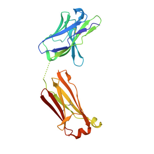
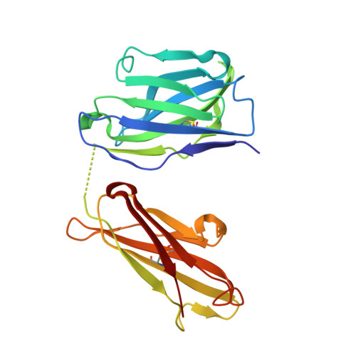
Highly specific anti-estradiol antibodies: structural characterisation and binding diversity.

Monnet, C.Bettsworth, F.Stura, E.A.Du, M.H.Menez, R.Derrien, L.Zinn-Justin, S.Gilquin, B.Sibai, G.Battail-Poirot, N.Jolivet, M.Menez, A.Arnaud, M.Ducancel, F.Charbonnier, J.B.

(2002) J Mol Biol 315: 699-712

  • DOI: https://doi.org/10.1006/jmbi.2001.5284
  • Primary Citation of Related Structures:  
    1JN6, 1JNH, 1JNL, 1JNN

  • PubMed Abstract: 

    Subtle modulation of antibody-binding properties by protein engineering often lies with an accurate structural and energetic description of how an antigen is recognised. Thus, with the intent to increase the affinity and add a bias in favour of natural estradiol compared with its chemically modified immunogen, we have determined the crystal structure of two anti-estradiol monoclonal antibodies, 10G6D6 and 17E12E5. Although generated against the same estradiol derivative, these antibodies share little sequence identity, which is reflected in dissimilar binding pockets and in different positioning of the steroid. In both antibodies the characteristic 17-hydroxyl group is buried deeply at the bottom of hydrophobic pockets and stabilised by hydrogen bonds. Apart from this similarity, the steroid is oriented differently in the respective binding pockets. The high specificity of both antibodies has been mapped out, and even closely related steroids show low cross-reactivity. The structural studies of the complex formed between 10G6D6 and 6-CMO-estradiol have identified contacts between the 6-CMO coupling linker and an arginine residue from the heavy chain CDR2 segment. This segment is now being targeted by random mutagenesis to select mutants with a preference for natural estradiol compared to the branched hapten.


  • Organizational Affiliation

    Département d'Ingénierie et d'Etude des Protéines, CEA, CE Saclay, Gif-sur-Yvette Cedex, 91191, France.


Macromolecules
Find similar proteins by:  (by identity cutoff)  |  3D Structure
Entity ID: 1
MoleculeChains Sequence LengthOrganismDetailsImage
monoclonal anti-estradiol 10G6D6 Fab light chain
A, C, E, G
212Mus musculusMutation(s): 0 
UniProt
Find proteins for P01843 (Mus musculus)
Explore P01843 
Go to UniProtKB:  P01843
Entity Groups  
Sequence Clusters30% Identity50% Identity70% Identity90% Identity95% Identity100% Identity
UniProt GroupP01843
Sequence Annotations
Expand
  • Reference Sequence
Find similar proteins by:  (by identity cutoff)  |  3D Structure
Entity ID: 2
MoleculeChains Sequence LengthOrganismDetailsImage
monoclonal anti-estradiol 10G6D6 Fab heavy chain
B, D, F, H
218Mus musculusMutation(s): 0 
Entity Groups  
Sequence Clusters30% Identity50% Identity70% Identity90% Identity95% Identity100% Identity
Sequence Annotations
Expand
  • Reference Sequence
Experimental Data & Validation

Experimental Data

  • Method: X-RAY DIFFRACTION
  • Resolution: 2.85 Å
  • R-Value Free: 0.253 
  • R-Value Work: 0.198 
  • Space Group: P 1 21 1
Unit Cell:
Length ( Å )Angle ( ˚ )
a = 70.2α = 90
b = 192.9β = 92.93
c = 71.12γ = 90
Software Package:
Software NamePurpose
DENZOdata reduction
SCALEPACKdata scaling
AMoREphasing
CNSrefinement

Structure Validation

View Full Validation Report


Ligand Structure Quality Assessment 


Entry History 

Revision History  (Full details and data files)

  • Version 1.0: 2002-02-06
    Type: Initial release
  • Version 1.1: 2008-04-27
    Changes: Version format compliance
  • Version 1.2: 2011-07-13
    Changes: Version format compliance
  • Version 1.3: 2017-06-28
    Changes: Database references, Source and taxonomy, Structure summary
  • Version 1.4: 2023-08-16
    Changes: Data collection, Database references, Derived calculations, Refinement description