1JMU

Crystal Structure of the Reovirus mu1/sigma3 Complex


Experimental Data Snapshot

  • Method: X-RAY DIFFRACTION
  • Resolution: 2.80 Å
  • R-Value Free: 0.241 
  • R-Value Work: 0.216 
  • R-Value Observed: 0.216 

wwPDB Validation   3D Report Full Report


Ligand Structure Quality Assessment 


This is version 1.4 of the entry. See complete history


Literature

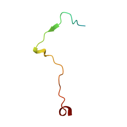
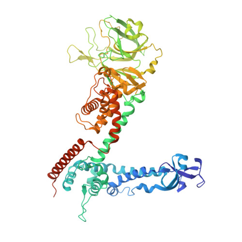
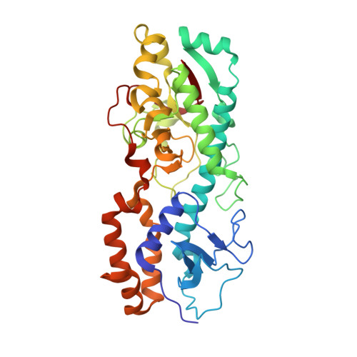
Structure of the reovirus membrane-penetration protein, Mu1, in a complex with is protector protein, Sigma3.

Liemann, S.Chandran, K.Baker, T.S.Nibert, M.L.Harrison, S.C.

(2002) Cell 108: 283-295

  • DOI: https://doi.org/10.1016/s0092-8674(02)00612-8
  • Primary Citation of Related Structures:  
    1JMU

  • PubMed Abstract: 

    Cell entry by nonenveloped animal viruses requires membrane penetration without membrane fusion. The reovirus penetration agent is the outer-capsid protein, Mu1. The structure of Mu1, complexed with its "protector" protein, Sigma3, and the fit of this Mu1(3)Sigma3(3) heterohexameric complex into the cryoEM image of an intact virion, reveal molecular events essential for viral penetration. Autolytic cleavage divides Mu1 into myristoylated Mu1N and Mu1C. A long hydrophobic pocket can receive the myristoyl group. Dissociation of Mu1N, linked to a major conformational change of the entire Mu1 trimer, must precede myristoyl-group insertion into the cellular membrane. A myristoyl switch, coupling exposure of the fatty acid chain, autolytic cleavage of Mu1N, and long-range molecular rearrangement of Mu1C, thus appears to be part of the penetration mechanism.


  • Organizational Affiliation

    Howard Hughes Medical Institute, Children's Hospital, Harvard Medical School, 320 Longwood Avenue, Boston, MA 02115, USA.


Macromolecules
Find similar proteins by:  (by identity cutoff)  |  3D Structure
Entity ID: 1
MoleculeChains Sequence LengthOrganismDetailsImage
PROTEIN MU-1
A, C, E
41Reovirus sp.Mutation(s): 0 
Gene Names: M2
UniProt
Find proteins for P11077 (Reovirus type 1 (strain Lang))
Explore P11077 
Go to UniProtKB:  P11077
Entity Groups  
Sequence Clusters30% Identity50% Identity70% Identity90% Identity95% Identity100% Identity
UniProt GroupP11077
Sequence Annotations
Expand
  • Reference Sequence
Find similar proteins by:  (by identity cutoff)  |  3D Structure
Entity ID: 2
MoleculeChains Sequence LengthOrganismDetailsImage
PROTEIN MU-1
B, D, F
666Reovirus sp.Mutation(s): 0 
Gene Names: M2
UniProt
Find proteins for P11077 (Reovirus type 1 (strain Lang))
Explore P11077 
Go to UniProtKB:  P11077
Entity Groups  
Sequence Clusters30% Identity50% Identity70% Identity90% Identity95% Identity100% Identity
UniProt GroupP11077
Sequence Annotations
Expand
  • Reference Sequence
Find similar proteins by:  (by identity cutoff)  |  3D Structure
Entity ID: 3
MoleculeChains Sequence LengthOrganismDetailsImage
SIGMA 3 PROTEIN
G, H, I
366Reovirus sp.Mutation(s): 1 
Gene Names: S4
UniProt
Find proteins for P07939 (Reovirus type 1 (strain Lang))
Explore P07939 
Go to UniProtKB:  P07939
Entity Groups  
Sequence Clusters30% Identity50% Identity70% Identity90% Identity95% Identity100% Identity
UniProt GroupP07939
Sequence Annotations
Expand
  • Reference Sequence
Small Molecules
Ligands 4 Unique
IDChains Name / Formula / InChI Key2D Diagram3D Interactions
BOG
Query on BOG

Download Ideal Coordinates CCD File 
K [auth B],
M [auth D],
O [auth F]
octyl beta-D-glucopyranoside
C14 H28 O6
HEGSGKPQLMEBJL-RKQHYHRCSA-N
SO4
Query on SO4

Download Ideal Coordinates CCD File 
L [auth B]
N [auth D]
P [auth F]
R [auth G]
S [auth G]
L [auth B],
N [auth D],
P [auth F],
R [auth G],
S [auth G],
U [auth H],
V [auth H],
X [auth I],
Y [auth I]
SULFATE ION
O4 S
QAOWNCQODCNURD-UHFFFAOYSA-L
ZN
Query on ZN

Download Ideal Coordinates CCD File 
Q [auth G],
T [auth H],
W [auth I]
ZINC ION
Zn
PTFCDOFLOPIGGS-UHFFFAOYSA-N
CL
Query on CL

Download Ideal Coordinates CCD File 
J [auth A]CHLORIDE ION
Cl
VEXZGXHMUGYJMC-UHFFFAOYSA-M
Experimental Data & Validation

Experimental Data

  • Method: X-RAY DIFFRACTION
  • Resolution: 2.80 Å
  • R-Value Free: 0.241 
  • R-Value Work: 0.216 
  • R-Value Observed: 0.216 
  • Space Group: I 2 2 2
Unit Cell:
Length ( Å )Angle ( ˚ )
a = 180.294α = 90
b = 184.942β = 90
c = 284.324γ = 90
Software Package:
Software NamePurpose
MLPHAREphasing
CNSrefinement
DENZOdata reduction
SCALEPACKdata scaling

Structure Validation

View Full Validation Report


Ligand Structure Quality Assessment 


Entry History 

Deposition Data

Revision History  (Full details and data files)

  • Version 1.0: 2002-02-06
    Type: Initial release
  • Version 1.1: 2008-04-27
    Changes: Version format compliance
  • Version 1.2: 2011-07-13
    Changes: Version format compliance
  • Version 1.3: 2020-07-29
    Type: Remediation
    Reason: Carbohydrate remediation
    Changes: Data collection, Derived calculations, Structure summary
  • Version 1.4: 2021-10-27
    Changes: Database references, Structure summary