1JL2

Crystal structure of TCEO RNase H-a chimera combining the folding core from T. thermophilus RNase H and the remaining region of E. coli RNase H


Experimental Data Snapshot

  • Method: X-RAY DIFFRACTION
  • Resolution: 1.76 Å
  • R-Value Free: 0.286 
  • R-Value Work: 0.249 

wwPDB Validation   3D Report Full Report


This is version 1.4 of the entry. See complete history


Literature

Contributions of folding cores to the thermostabilities of two ribonucleases H.

Robic, S.Berger, J.M.Marqusee, S.

(2002) Protein Sci 11: 381-389

  • DOI: https://doi.org/10.1110/ps.38602
  • Primary Citation of Related Structures:  
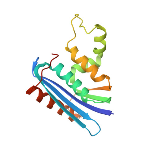
    1JL2

  • PubMed Abstract: 

    To investigate the contribution of the folding cores to the thermodynamic stability of RNases H, we used rational design to create two chimeras composed of parts of a thermophilic and a mesophilic RNase H. Each chimera combines the folding core from one parent protein and the remaining parts of the other. Both chimeras form active, well-folded RNases H. Stability curves, based on CD-monitored chemical denaturations, show that the chimera with the thermophilic core is more stable, has a higher midpoint of thermal denaturation, and a lower change in heat capacity (DeltaCp) upon unfolding than the chimera with the mesophilic core. A possible explanation for the low DeltaCp of both the parent thermophilic RNase H and the chimera with the thermophilic core is the residual structure in the denatured state. On the basis of the studied parameters, the chimera with the thermophilic core resembles a true thermophilic protein. Our results suggest that the folding core plays an essential role in conferring thermodynamic parameters to RNases H.


  • Organizational Affiliation

    Department of Molecular and Cell Biology, University of California, Berkeley, California 94720, USA.


Macromolecules
Find similar proteins by:  (by identity cutoff)  |  3D Structure
Entity ID: 1
MoleculeChains Sequence LengthOrganismDetailsImage
Chimera of Ribonuclease HI, Ribonuclease H
A, B, C, D
156Escherichia coli K-12Thermus thermophilus HB8Mutation(s): 0 
Gene Names: rnhAdasFherArnhsdrAb0214JW0204TTHA1556
EC: 3.1.26.4
UniProt
Find proteins for P0A7Y4 (Escherichia coli (strain K12))
Explore P0A7Y4 
Go to UniProtKB:  P0A7Y4
Find proteins for P29253 (Thermus thermophilus (strain ATCC 27634 / DSM 579 / HB8))
Explore P29253 
Go to UniProtKB:  P29253
Entity Groups  
Sequence Clusters30% Identity50% Identity70% Identity90% Identity95% Identity100% Identity
UniProt GroupsP0A7Y4P29253
Sequence Annotations
Expand
  • Reference Sequence
Experimental Data & Validation

Experimental Data

  • Method: X-RAY DIFFRACTION
  • Resolution: 1.76 Å
  • R-Value Free: 0.286 
  • R-Value Work: 0.249 
  • Space Group: P 1 21 1
Unit Cell:
Length ( Å )Angle ( ˚ )
a = 56.195α = 90
b = 87.033β = 91.87
c = 60.687γ = 90
Software Package:
Software NamePurpose
CNSrefinement
DENZOdata reduction
SCALEPACKdata scaling
CNSphasing

Structure Validation

View Full Validation Report


Entry History 

Deposition Data

Revision History  (Full details and data files)

  • Version 1.0: 2002-01-18
    Type: Initial release
  • Version 1.1: 2008-04-27
    Changes: Version format compliance
  • Version 1.2: 2011-07-13
    Changes: Version format compliance
  • Version 1.3: 2017-06-28
    Changes: Database references, Source and taxonomy, Structure summary
  • Version 1.4: 2023-08-16
    Changes: Data collection, Database references, Refinement description