1JDR

Crystal Structure of a Proximal Domain Potassium Binding Variant of Cytochrome c Peroxidase


Experimental Data Snapshot

  • Method: X-RAY DIFFRACTION
  • Resolution: 1.50 Å
  • R-Value Free: 0.234 
  • R-Value Work: 0.197 

wwPDB Validation   3D Report Full Report


Ligand Structure Quality Assessment 


This is version 1.5 of the entry. See complete history


Literature

The effects of an engineered cation site on the structure, activity, and EPR properties of cytochrome c peroxidase.

Bonagura, C.A.Sundaramoorthy, M.Bhaskar, B.Poulos, T.L.

(1999) Biochemistry 38: 5538-5545

  • DOI: https://doi.org/10.1021/bi982996k
  • Primary Citation of Related Structures:  
    1JDR

  • PubMed Abstract: 

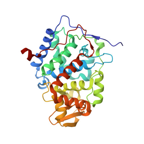
    Earlier work [Bonagura et al. (1996) Biochemistry 35, 6107] showed that the K+ site found in the proximal pocket of ascorbate peroxidase (APX) could be engineered into cytochrome c peroxidase (CCP). Binding of K+ at the engineered site results in a loss in activity and destabilization of the CCP compound I Trp191 cationic radical owing to long-range electrostatic effects. The engineered CCP mutant crystal structure has been refined to 1.5 A using data obtained at cryogenic temperatures which provides a more detailed basis for comparison with the naturally occurring K+ site in APX. The characteristic EPR signal associated with the Trp191 radical becomes progressively weaker as K+ is added, which correlates well with the loss in enzyme activity as [K+] is increased. These results coupled with stopped-flow studies support our earlier conclusions that the loss in activity and EPR signal is due to destabilization of the Trp191 cationic radical.


  • Organizational Affiliation

    Department of Molecular Biology & Biochemistry, University of California, Irvine 92697-3900, USA.


Macromolecules
Find similar proteins by:  (by identity cutoff)  |  3D Structure
Entity ID: 1
MoleculeChains Sequence LengthOrganismDetailsImage
Cytochrome c Peroxidase294Saccharomyces cerevisiaeMutation(s): 5 
Gene Names: OPBYC
EC: 1.11.1.5
UniProt
Find proteins for P00431 (Saccharomyces cerevisiae (strain ATCC 204508 / S288c))
Explore P00431 
Go to UniProtKB:  P00431
Entity Groups  
Sequence Clusters30% Identity50% Identity70% Identity90% Identity95% Identity100% Identity
UniProt GroupP00431
Sequence Annotations
Expand
  • Reference Sequence
Small Molecules
Ligands 2 Unique
IDChains Name / Formula / InChI Key2D Diagram3D Interactions
HEM
Query on HEM

Download Ideal Coordinates CCD File 
C [auth A]PROTOPORPHYRIN IX CONTAINING FE
C34 H32 Fe N4 O4
KABFMIBPWCXCRK-RGGAHWMASA-L
K
Query on K

Download Ideal Coordinates CCD File 
B [auth A]POTASSIUM ION
K
NPYPAHLBTDXSSS-UHFFFAOYSA-N
Experimental Data & Validation

Experimental Data

  • Method: X-RAY DIFFRACTION
  • Resolution: 1.50 Å
  • R-Value Free: 0.234 
  • R-Value Work: 0.197 
  • Space Group: P 21 21 21
Unit Cell:
Length ( Å )Angle ( ˚ )
a = 106.717α = 90
b = 75.531β = 90
c = 51.256γ = 90
Software Package:
Software NamePurpose
AMoREphasing
X-PLORrefinement
MOSFLMdata reduction
SCALEPACKdata scaling

Structure Validation

View Full Validation Report


Ligand Structure Quality Assessment 


Entry History 

Deposition Data

Revision History  (Full details and data files)

  • Version 1.0: 2001-06-27
    Type: Initial release
  • Version 1.1: 2008-04-27
    Changes: Version format compliance
  • Version 1.2: 2011-07-13
    Changes: Version format compliance
  • Version 1.3: 2021-10-27
    Changes: Database references, Derived calculations
  • Version 1.4: 2024-02-07
    Changes: Data collection
  • Version 1.5: 2024-04-03
    Changes: Refinement description