1J71

Structure of the extracellular aspartic proteinase from Candida tropicalis yeast.


Experimental Data Snapshot

  • Method: X-RAY DIFFRACTION
  • Resolution: 1.80 Å
  • R-Value Free: 0.193 
  • R-Value Work: 0.165 
  • R-Value Observed: 0.168 

wwPDB Validation   3D Report Full Report


This is version 1.4 of the entry. See complete history


Literature

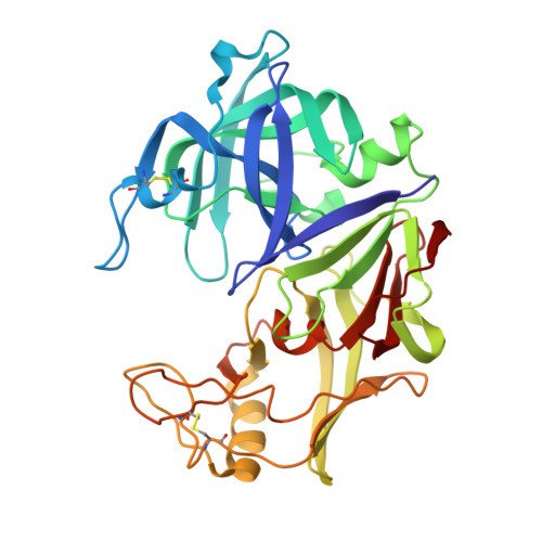
High-resolution structure of the extracellular aspartic proteinase from Candida tropicalis yeast.

Symersky, J.Monod, M.Foundling, S.I.

(1997) Biochemistry 36: 12700-12710

  • DOI: https://doi.org/10.1021/bi970613x
  • Primary Citation of Related Structures:  
    1J71

  • PubMed Abstract: 

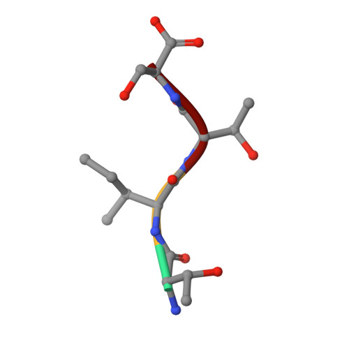
    The crystal structure of the secreted aspartic proteinase from Candida tropicalis yeast (SAPT) has been determined to 1.8 A resolution. The classic aspartic proteinase bilobal structure and domain topology is conserved in SAPT, with the substrate binding cleft situated between the two domains. Structural comparisons made with pepsin indicate that insertions and deletions in the primary sequence modify the SAPT structure to create a more spacious substrate binding cleft with altered specificity. An unexpected tetrapeptide has been found to occupy binding sites S1'-S3', and this suggests the order of release of peptide products in the catalytic mechanism of these enzymes. Structural features are considered with regard to previous substrate specificity data.


  • Organizational Affiliation

    Laboratory of Protein Crystallography, Crystallography Research Program, Oklahoma Medical Research Foundation, 825 N.E. 13th Street, Oklahoma City, Oklahoma 73104, USA.


Macromolecules
Find similar proteins by:  (by identity cutoff)  |  3D Structure
Entity ID: 1
MoleculeChains Sequence LengthOrganismDetailsImage
Aspartic proteinase334Candida tropicalisMutation(s): 0 
EC: 3.4.23.24
UniProt
Find proteins for Q00663 (Candida tropicalis)
Explore Q00663 
Go to UniProtKB:  Q00663
Entity Groups  
Sequence Clusters30% Identity50% Identity70% Identity90% Identity95% Identity100% Identity
UniProt GroupQ00663
Sequence Annotations
Expand
  • Reference Sequence

Find similar proteins by:  Sequence   |   3D Structure  

Entity ID: 2
MoleculeChains Sequence LengthOrganismDetailsImage
Tetrapeptide Thr-Ile-Thr-Ser4unidentifiedMutation(s): 0 
Sequence Annotations
Expand
  • Reference Sequence
Experimental Data & Validation

Experimental Data

  • Method: X-RAY DIFFRACTION
  • Resolution: 1.80 Å
  • R-Value Free: 0.193 
  • R-Value Work: 0.165 
  • R-Value Observed: 0.168 
  • Space Group: P 21 21 21
Unit Cell:
Length ( Å )Angle ( ˚ )
a = 49.96α = 90
b = 51.43β = 90
c = 128.9γ = 90
Software Package:
Software NamePurpose
FRAMBOdata collection
SAINTdata reduction
AMoREphasing
CNSrefinement
SAINTdata scaling

Structure Validation

View Full Validation Report


Entry History 

Deposition Data

Revision History  (Full details and data files)

  • Version 1.0: 2001-05-23
    Type: Initial release
  • Version 1.1: 2008-04-27
    Changes: Version format compliance
  • Version 1.2: 2011-07-13
    Changes: Version format compliance
  • Version 1.3: 2017-10-04
    Changes: Refinement description
  • Version 1.4: 2023-08-16
    Changes: Data collection, Database references, Derived calculations, Refinement description