1IXE

Crystal structure of citrate synthase from Thermus thermophilus HB8


Experimental Data Snapshot

  • Method: X-RAY DIFFRACTION
  • Resolution: 2.30 Å
  • R-Value Free: 0.217 
  • R-Value Work: 0.177 
  • R-Value Observed: 0.179 

wwPDB Validation   3D Report Full Report


Ligand Structure Quality Assessment 


This is version 1.6 of the entry. See complete history


Literature

Structural comparison between the open and closed forms of citrate synthase from Thermus thermophilus HB8.

Kanamori, E.Kawaguchi, S.Kuramitsu, S.Kouyama, T.Murakami, M.

(2015) Biophys Physicobiol 12: 47-56

  • DOI: https://doi.org/10.2142/biophysico.12.0_47
  • Primary Citation of Related Structures:  
    1IOM, 1IXE

  • PubMed Abstract: 

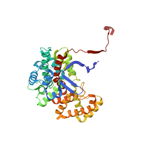
    The crystal structures of citrate synthase from the thermophilic eubacteria Thermus thermophilus HB8 (TtCS) were determined for an open form at 1.5 Å resolution and for closed form at 2.3 Å resolution, respectively. In the absence of ligands TtCS in the open form was crystalized into a tetragonal form with a single subunit in the asymmetric unit. TtCS was also co-crystallized with citrate and coenzyme-A to form an orthorhombic crystal with two homodimers in the asymmetric unit. Citrate and CoA are found in the active site situated between the large domain and the small domain in all subunit whereas the complex shows two distinct closed conformations, the fully closed form and partially closed form. Structural comparisons are performed to describe conformational changes associated with binding of products of TtCS. Upon binding of citrate, basic residues in the active site move toward citrate and make a hydrogen bond network in the active site, inducing a large-scale rotation of the small domain relative to the large domain. CoA is sandwiched between the small and large domains and then the cysteamine tail is inserted into the active site with a cooperative rotation around mainchain dihedrals in the hinge region connecting helices M and N. According to this rotation these helices are extended to close the active site completely. The considerable flexibility and structural rearrangements in the hinge region are crucial for an ordered bibi reaction in catalysis for microbial CSs.


  • Organizational Affiliation

    Department of Physics, Graduate School of Science, Nagoya University, Nagoya, Aichi 464-8602, Japan.


Macromolecules
Find similar proteins by:  (by identity cutoff)  |  3D Structure
Entity ID: 1
MoleculeChains Sequence LengthOrganismDetailsImage
citrate synthase
A, B, C, D
377Thermus thermophilusMutation(s): 0 
Gene Names: HB8
EC: 4.1.3.7
UniProt
Find proteins for Q5SIM6 (Thermus thermophilus (strain ATCC 27634 / DSM 579 / HB8))
Explore Q5SIM6 
Go to UniProtKB:  Q5SIM6
Entity Groups  
Sequence Clusters30% Identity50% Identity70% Identity90% Identity95% Identity100% Identity
UniProt GroupQ5SIM6
Sequence Annotations
Expand
  • Reference Sequence
Small Molecules
Ligands 4 Unique
IDChains Name / Formula / InChI Key2D Diagram3D Interactions
COA
Query on COA

Download Ideal Coordinates CCD File 
G [auth A],
M [auth B],
S [auth C],
X [auth D]
COENZYME A
C21 H36 N7 O16 P3 S
RGJOEKWQDUBAIZ-IBOSZNHHSA-N
CIT
Query on CIT

Download Ideal Coordinates CCD File 
H [auth A],
N [auth B],
T [auth C],
Y [auth D]
CITRIC ACID
C6 H8 O7
KRKNYBCHXYNGOX-UHFFFAOYSA-N
SO4
Query on SO4

Download Ideal Coordinates CCD File 
E [auth A]
F [auth A]
J [auth B]
K [auth B]
L [auth B]
E [auth A],
F [auth A],
J [auth B],
K [auth B],
L [auth B],
P [auth C],
Q [auth C],
R [auth C],
V [auth D],
W [auth D]
SULFATE ION
O4 S
QAOWNCQODCNURD-UHFFFAOYSA-L
GOL
Query on GOL

Download Ideal Coordinates CCD File 
I [auth A],
O [auth B],
U [auth C],
Z [auth D]
GLYCEROL
C3 H8 O3
PEDCQBHIVMGVHV-UHFFFAOYSA-N
Experimental Data & Validation

Experimental Data

  • Method: X-RAY DIFFRACTION
  • Resolution: 2.30 Å
  • R-Value Free: 0.217 
  • R-Value Work: 0.177 
  • R-Value Observed: 0.179 
  • Space Group: P 21 21 21
Unit Cell:
Length ( Å )Angle ( ˚ )
a = 80.01α = 90
b = 110.63β = 90
c = 184.382γ = 90
Software Package:
Software NamePurpose
MOSFLMdata reduction
SCALAdata scaling
CNSrefinement
CCP4data scaling
CNSphasing

Structure Validation

View Full Validation Report


Ligand Structure Quality Assessment 


Entry History 

Revision History  (Full details and data files)

  • Version 1.0: 2003-07-29
    Type: Initial release
  • Version 1.1: 2008-04-27
    Changes: Version format compliance
  • Version 1.2: 2011-07-13
    Changes: Non-polymer description, Version format compliance
  • Version 1.3: 2014-04-16
    Changes: Other
  • Version 1.4: 2015-11-04
    Changes: Database references
  • Version 1.5: 2020-01-01
    Changes: Database references
  • Version 1.6: 2023-10-25
    Changes: Data collection, Database references, Derived calculations, Refinement description