1IWQ

Crystal Structure of MARCKS calmodulin binding domain peptide complexed with Ca2+/Calmodulin


Experimental Data Snapshot

  • Method: X-RAY DIFFRACTION
  • Resolution: 2.00 Å
  • R-Value Free: 0.267 
  • R-Value Work: 0.223 
  • R-Value Observed: 0.225 

wwPDB Validation   3D Report Full Report


This is version 1.3 of the entry. See complete history


Literature

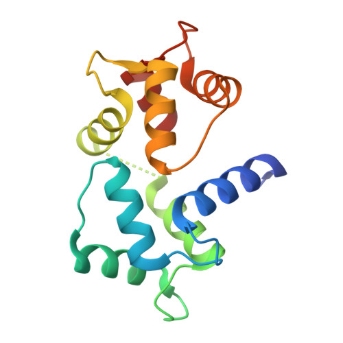
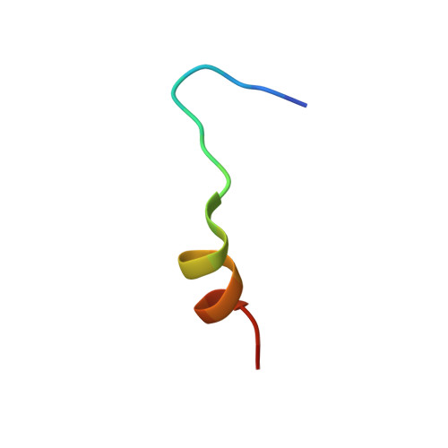
Crystal structure of a MARCKS peptide containing the calmodulin-binding domain in complex with Ca(2+)-calmodulin

Yamauchi, E.Nakatsu, T.Matsubara, M.Kato, H.Taniguchi, H.

(2003) Nat Struct Biol 10: 226-231

  • DOI: https://doi.org/10.1038/nsb900
  • Primary Citation of Related Structures:  
    1IWQ

  • PubMed Abstract: 

    The calmodulin-binding domain of myristoylated alanine-rich C kinase substrate (MARCKS), which interacts with various targets including calmodulin, actin and membrane lipids, has been suggested to function as a crosstalk point among several signal transduction pathways. We present here the crystal structure at 2 A resolution of a peptide consisting of the MARCKS calmodulin (CaM)-binding domain in complex with Ca2+-CaM. The domain assumes a flexible conformation, and the hydrophobic pocket of the calmodulin N-lobe, which is a common CaM-binding site observed in previously resolved Ca2+-CaM-target peptide complexes, is not involved in the interaction. The present structure presents a novel target-recognition mode of calmodulin and provides insight into the structural basis of the flexible interaction module of MARCKS.


  • Organizational Affiliation

    Harima Institute at SPring-8, RIKEN, 1-1-1 Kouto, Sayo, Hyogo 679-5148, Japan.


Macromolecules
Find similar proteins by:  (by identity cutoff)  |  3D Structure
Entity ID: 1
MoleculeChains Sequence LengthOrganismDetailsImage
CALMODULIN148Homo sapiensMutation(s): 0 
UniProt & NIH Common Fund Data Resources
Find proteins for P0DP23 (Homo sapiens)
Explore P0DP23 
Go to UniProtKB:  P0DP23
PHAROS:  P0DP23
GTEx:  ENSG00000198668 
Entity Groups  
Sequence Clusters30% Identity50% Identity70% Identity90% Identity95% Identity100% Identity
UniProt GroupP0DP23
Sequence Annotations
Expand
  • Reference Sequence

Find similar proteins by:  Sequence   |   3D Structure  

Entity ID: 2
MoleculeChains Sequence LengthOrganismDetailsImage
MARCKS19N/AMutation(s): 0 
UniProt & NIH Common Fund Data Resources
Find proteins for P29966 (Homo sapiens)
Explore P29966 
Go to UniProtKB:  P29966
PHAROS:  P29966
GTEx:  ENSG00000277443 
Entity Groups  
Sequence Clusters30% Identity50% Identity70% Identity90% Identity95% Identity100% Identity
UniProt GroupP29966
Sequence Annotations
Expand
  • Reference Sequence
Experimental Data & Validation

Experimental Data

  • Method: X-RAY DIFFRACTION
  • Resolution: 2.00 Å
  • R-Value Free: 0.267 
  • R-Value Work: 0.223 
  • R-Value Observed: 0.225 
  • Space Group: P 61 2 2
Unit Cell:
Length ( Å )Angle ( ˚ )
a = 40.229α = 90
b = 40.229β = 90
c = 343.468γ = 120
Software Package:
Software NamePurpose
DENZOdata reduction
SCALEPACKdata scaling
AMoREphasing
REFMACrefinement

Structure Validation

View Full Validation Report


Entry History 

Deposition Data

Revision History  (Full details and data files)

  • Version 1.0: 2003-03-11
    Type: Initial release
  • Version 1.1: 2008-04-27
    Changes: Version format compliance
  • Version 1.2: 2011-07-13
    Changes: Version format compliance
  • Version 1.3: 2023-10-25
    Changes: Data collection, Database references, Derived calculations, Refinement description