1IVR

STRUCTURE OF ASPARTATE AMINOTRANSFERASE


Experimental Data Snapshot

  • Method: X-RAY DIFFRACTION
  • Resolution: 2.40 Å
  • R-Value Work: 0.153 

wwPDB Validation   3D Report Full Report


This is version 1.6 of the entry. See complete history


Literature

Aspartate aminotransferase complexed with erythro-beta-hydroxyaspartate: crystallographic and spectroscopic identification of the carbinolamine intermediate.

von Stosch, A.G.

(1996) Biochemistry 35: 15260-15268

  • DOI: https://doi.org/10.1021/bi960994z
  • Primary Citation of Related Structures:  
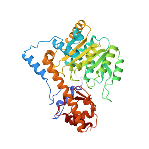
    1IVR

  • PubMed Abstract: 

    The crystal structure of mitochondrial aspartate aminotransferase (mAAT) of chicken complexed with erythro-beta-hydroxyaspartate has been determined at 2.4 A resolution. Pregrown crystals of mAAT complexed with the inhibitor maleate (closed enzyme conformation, orthorhombic space group C222(1)) were soaked in solutions of erythro-beta-hydroxyaspartate. The ligand exchange was monitored by microspectrophotometry. The active site turned out to be predominantly occupied by the carbinolamine intermediate. The carbinolamine is a true intermediate of the catalytic cycle forming the last covalently bound enzyme:substrate complex before release of the keto acid product. Occupancies of approximately 80% for the carbinolamine and of approximately 20% for the quinonoid intermediate were obtained. Two hydrogen bonds were identified that are potentially relevant for the accumulation of the carbinolamine intermediate: one to the hydroxyl group of Tyr 70* and the other to the epsilon-NH2 group of Lys 258.


Macromolecules
Find similar proteins by:  (by identity cutoff)  |  3D Structure
Entity ID: 1
MoleculeChains Sequence LengthOrganismDetailsImage
ASPARTATE AMINOTRANSFERASE401Gallus gallusMutation(s): 0 
EC: 2.6.1.1
UniProt
Find proteins for P00508 (Gallus gallus)
Explore P00508 
Go to UniProtKB:  P00508
Entity Groups  
Sequence Clusters30% Identity50% Identity70% Identity90% Identity95% Identity100% Identity
UniProt GroupP00508
Sequence Annotations
Expand
  • Reference Sequence
Small Molecules
Ligands 1 Unique
IDChains Name / Formula / InChI Key2D Diagram3D Interactions
CBA
Query on CBA

Download Ideal Coordinates CCD File 
B [auth A]N-PYRIDOXYL-2,3-DIHYDROXYASPARTIC ACID-5-MONOPHOSPHATE
C12 H17 N2 O11 P
SMJZVFGOJYUMBO-JOYOIKCWSA-N
Experimental Data & Validation

Experimental Data

  • Method: X-RAY DIFFRACTION
  • Resolution: 2.40 Å
  • R-Value Work: 0.153 
  • Space Group: C 2 2 21
Unit Cell:
Length ( Å )Angle ( ˚ )
a = 69.7α = 90
b = 91.4β = 90
c = 128.5γ = 90
Software Package:
Software NamePurpose
TNTrefinement
PROLSQrefinement

Structure Validation

View Full Validation Report


Entry History 

Deposition Data

Revision History  (Full details and data files)

  • Version 1.0: 1997-07-23
    Type: Initial release
  • Version 1.1: 2008-03-24
    Changes: Version format compliance
  • Version 1.2: 2011-07-13
    Changes: Derived calculations, Version format compliance
  • Version 1.3: 2018-04-18
    Changes: Data collection, Other
  • Version 1.4: 2019-07-17
    Changes: Data collection, Refinement description
  • Version 1.5: 2019-08-14
    Changes: Data collection
  • Version 1.6: 2024-02-07
    Changes: Data collection, Database references, Derived calculations