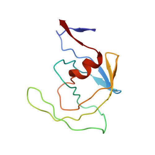
1IVP

THE CRYSTALLOGRAPHIC STRUCTURE OF THE PROTEASE FROM HUMAN IMMUNODEFICIENCY VIRUS TYPE 2 WITH TWO SYNTHETIC PEPTIDIC TRANSITION STATE ANALOG INHIBITORS


Experimental Data Snapshot

  • Method: X-RAY DIFFRACTION
  • Resolution: 2.50 Å
  • R-Value Observed: 0.197 

wwPDB Validation   3D Report Full Report


This is version 1.5 of the entry. See complete history


Literature

The crystallographic structure of the protease from human immunodeficiency virus type 2 with two synthetic peptidic transition state analog inhibitors.

Mulichak, A.M.Hui, J.O.Tomasselli, A.G.Heinrikson, R.L.Curry, K.A.Tomich, C.S.Thaisrivongs, S.Sawyer, T.K.Watenpaugh, K.D.

(1993) J Biol Chem 268: 13103-13109

  • Primary Citation of Related Structures:  
    1IVP, 1IVQ

  • PubMed Abstract: 

    The crystal structure of human immunodeficiency virus (HIV) type 2 protease has been determined in complexes with peptidic inhibitors Noa-His-Cha psi [CH(OH)CH(OH)]Val-Ile-Amp (U75875) and Qnc-Asn-Cha psi [CH(OH)CH2]Val-Npt(U92163) (where Noa is naphthyloxyacetyl, Cha is cyclohexylalanine, Amp is 2-aminomethylpyridine, Qnc is quinoline-2-carbonyl, and Npt is neopentylamine), which have dihydroxyethylene and hydroxyethylene moieties, respectively, in place of the normal scissile bond of the natural ligand. The complexes crystallize in space group P2(1)2(1)2(1), with one dimer-inhibitor complex per asymmetric unit and average cell dimensions of a = 33.28 A, b = 45.35 A, c = 135.84 A. Data were collected to approximately 2.5-A resolution. The model structures were refined with resulting R-factors of around 0.19. As expected, the HIV-2 protease structure is approximately C2-symmetric with a gross structure very similar to that of the HIV-1 enzyme. The inhibitors bind in an extended conformation positioned lengthwise in the binding cleft in a manner similar to that found in the HIV-1 protease-inhibitor complexes previously reported. The substitution of the bulkier Ile82 side chain in the HIV-2 protease may help explain the better ability of HIV-2 protease to bind and hydrolyze ligands with small P1 and P1' side groups. It appears that differences in specificity between the proteases of HIV-1 and HIV-2 are not merely a result of simple side chain substitutions, but may be complicated by differences in main chain flexibility as well.


  • Organizational Affiliation

    Discovery Research, Upjohn Company, Kalamazoo, Michigan 49007.


Macromolecules
Find similar proteins by:  (by identity cutoff)  |  3D Structure
Entity ID: 1
MoleculeChains Sequence LengthOrganismDetailsImage
HIV-2 PROTEASE
A, B
99Human immunodeficiency virus 2Mutation(s): 0 
UniProt
Find proteins for P04584 (Human immunodeficiency virus type 2 subtype A (isolate ROD))
Explore P04584 
Go to UniProtKB:  P04584
Entity Groups  
Sequence Clusters30% Identity50% Identity70% Identity90% Identity95% Identity100% Identity
UniProt GroupP04584
Sequence Annotations
Expand
  • Reference Sequence
Small Molecules
Ligands 1 Unique
IDChains Name / Formula / InChI Key2D Diagram3D Interactions
1ZK
Query on 1ZK

Download Ideal Coordinates CCD File 
C [auth B]4-[(2R)-3-{[(1S,2S,3R,4S)-1-(cyclohexylmethyl)-2,3-dihydroxy-5-methyl-4-({(1S,2R)-2-methyl-1-[(pyridin-2-ylmethyl)carba moyl]butyl}carbamoyl)hexyl]amino}-2-{[(naphthalen-1-yloxy)acetyl]amino}-3-oxopropyl]-1H-imidazol-3-ium
C45 H62 N7 O7
ZTKCGLWIXHAITR-JIFWBGJESA-O
Binding Affinity Annotations 
IDSourceBinding Affinity
1ZK Binding MOAD:  1IVP Ki: 30 (nM) from 1 assay(s)
Biologically Interesting Molecules (External Reference) 1 Unique
Experimental Data & Validation

Experimental Data

  • Method: X-RAY DIFFRACTION
  • Resolution: 2.50 Å
  • R-Value Observed: 0.197 
  • Space Group: P 21 21 21
Unit Cell:
Length ( Å )Angle ( ˚ )
a = 33.19α = 90
b = 44.95β = 90
c = 135.65γ = 90
Software Package:
Software NamePurpose
PROLSQrefinement

Structure Validation

View Full Validation Report


Entry History 

Deposition Data

Revision History  (Full details and data files)

  • Version 1.0: 1993-07-15
    Type: Initial release
  • Version 1.1: 2008-03-24
    Changes: Version format compliance
  • Version 1.2: 2011-07-13
    Changes: Atomic model, Database references, Derived calculations, Non-polymer description, Structure summary, Version format compliance
  • Version 1.3: 2012-12-12
    Changes: Other
  • Version 1.4: 2017-11-29
    Changes: Derived calculations, Other
  • Version 1.5: 2024-02-07
    Changes: Data collection, Database references, Derived calculations, Structure summary