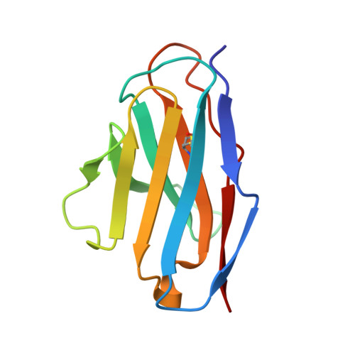
The de novo design of an antibody combining site. Crystallographic analysis of the VL domain confirms the structural model.
Essen, L.O., Skerra, A.(1994) J Mol Biol 238: 226-244
- PubMed: 8158652
- DOI: https://doi.org/10.1006/jmbi.1994.1284
- Primary Citation of Related Structures:
1IVL - PubMed Abstract:
In a protein design approach the molecular model of an artificial antibody Fv fragment was generated with predicted complementarity to part of the known crystal structure of chicken egg-white cystatin. The model of the Fv fragment was based on the three-dimensional structure of the anti-lysozyme antibody HyHEL-10, which was modified by substituting amino acid side-chains in the complementarity-determining regions (CDRs) as well as the framework without altering the backbone. In the course of crystallization experiments with the bacterially produced Fv fragment crystals of the VL domain alone were obtained. These crystals diffracted X-rays to a resolution of 2.17 A and were shown to belong to the space group P2(1)2(1)2(1) with unit cell dimensions a = 46.89 A, b = 58.05 A, c = 83.22 A containing two VL monomers in the asymmetric unit. The crystal structure was solved by molecular replacement and refined to a crystallographic R-factor of 17.5%. The two VL monomers exhibit an asymmetric mode of association, which is different from other crystallized VL domains described before and shows the peculiar feature of an isopropanol precipitant molecule buried at the interface. Both VL structures reveal a high level of similarity to the predicted three-dimensional model. With the exception of two loop segments in the framework region that are involved in crystal packing contacts, the backbone structures of the two VL monomers in the crystal and the molecular model of the VL domain are practically identical. Although six amino acid residues had been replaced in the hypervariable regions, the CDR conformations remained conserved and only minor deviations in the orientation of some side-chains and peptide planes were detected. The crystallographic analysis of the VL domain modelled as part of a complex between an artificial Fv fragment and the small protein cystatin, deliberately chosen as antigen target, confirms the concept of distinct structural classes for CDR backbones and supports our strategy for the de novo design of an antibody combining site.
Organizational Affiliation:
Max-Planck-Institut für Biophysik, Frankfurt am Main, Germany.