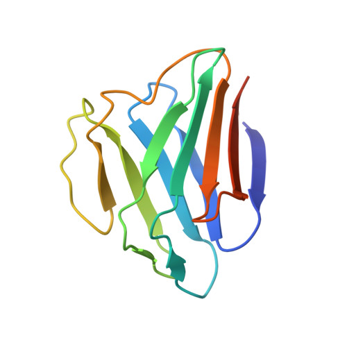
1IFR

Structure of Lamin A/C Globular Domain


Experimental Data Snapshot

  • Method: X-RAY DIFFRACTION
  • Resolution: 1.40 Å
  • R-Value Work: 0.216 

wwPDB Validation   3D Report Full Report


This is version 1.3 of the entry. See complete history


Literature

Structure of the globular tail of nuclear lamin.

Dhe-Paganon, S.Werner, E.D.Chi, Y.I.Shoelson, S.E.

(2002) J Biol Chem 277: 17381-17384

  • DOI: https://doi.org/10.1074/jbc.C200038200
  • Primary Citation of Related Structures:  
    1IFR

  • PubMed Abstract: 

    The nuclear lamins form a two-dimensional matrix that provides integrity to the cell nucleus and participates in nuclear activities. Mutations in the region of human LMNA encoding the carboxyl-terminal tail Lamin A/C are associated with forms of muscular dystrophy and familial partial lipodystrophy (FPLD). To help discriminate tissue-specific phenotypes, we have solved at 1.4-A resolution the three-dimensional crystal structure of the lamin A/C globular tail. The domain adopts a novel, all beta immunoglobulin-like fold. FPLD-associated mutations cluster within a small surface, whereas muscular dystrophy-associated mutations are distributed throughout the protein core and on its surface. These findings distinguish myopathy- and lipodystrophy-associated mutations and provide a structural framework for further testing hypotheses concerning lamin function.


  • Organizational Affiliation

    Joslin Diabetes Center & Department of Medicine, Harvard Medical School, Boston, Massachusetts 02215, USA.


Macromolecules
Find similar proteins by:  (by identity cutoff)  |  3D Structure
Entity ID: 1
MoleculeChains Sequence LengthOrganismDetailsImage
Lamin A/C121Homo sapiensMutation(s): 0 
UniProt & NIH Common Fund Data Resources
Find proteins for P02545 (Homo sapiens)
Explore P02545 
Go to UniProtKB:  P02545
PHAROS:  P02545
GTEx:  ENSG00000160789 
Entity Groups  
Sequence Clusters30% Identity50% Identity70% Identity90% Identity95% Identity100% Identity
UniProt GroupP02545
Sequence Annotations
Expand
  • Reference Sequence
Small Molecules
Ligands 1 Unique
IDChains Name / Formula / InChI Key2D Diagram3D Interactions
GOL
Query on GOL

Download Ideal Coordinates CCD File 
B [auth A]GLYCEROL
C3 H8 O3
PEDCQBHIVMGVHV-UHFFFAOYSA-N
Experimental Data & Validation

Experimental Data

  • Method: X-RAY DIFFRACTION
  • Resolution: 1.40 Å
  • R-Value Work: 0.216 
  • Space Group: P 21 21 21
Unit Cell:
Length ( Å )Angle ( ˚ )
a = 26.43α = 90
b = 52.55β = 90
c = 67.44γ = 90
Software Package:
Software NamePurpose
CNSrefinement

Structure Validation

View Full Validation Report


Entry History 

Deposition Data

Revision History  (Full details and data files)

  • Version 1.0: 2002-07-17
    Type: Initial release
  • Version 1.1: 2008-04-27
    Changes: Version format compliance
  • Version 1.2: 2011-07-13
    Changes: Non-polymer description, Version format compliance
  • Version 1.3: 2024-02-07
    Changes: Data collection, Database references, Derived calculations