1IBQ

ASPERGILLOPEPSIN FROM ASPERGILLUS PHOENICIS


Experimental Data Snapshot

  • Method: X-RAY DIFFRACTION
  • Resolution: 2.14 Å
  • R-Value Free: 0.269 
  • R-Value Work: 0.221 
  • R-Value Observed: 0.210 

wwPDB Validation   3D Report Full Report


Ligand Structure Quality Assessment 


This is version 1.4 of the entry. See complete history


Literature

Structure of aspergillopepsin I from Aspergillus phoenicis: variations of the S1'-S2 subsite in aspartic proteinases.

Cho, S.W.Kim, N.Choi, M.U.Shin, W.

(2001) Acta Crystallogr D Biol Crystallogr 57: 948-956

  • DOI: https://doi.org/10.1107/s0907444901005972
  • Primary Citation of Related Structures:  
    1IBQ

  • PubMed Abstract: 

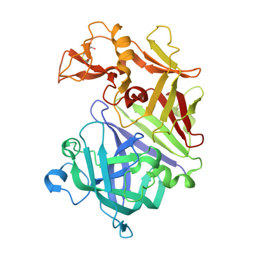
    The crystal structure of aspergillopepsin I (AP) from Aspergillus phoenicis has been determined at 2.18 A resolution and refined to R and R(free) factors of 21.5 and 26.0%, respectively. AP has the typical two beta-barrel domain structure of aspartic proteinases. The structures of the two independent molecules are partly different, exemplifying the flexible nature of the aspartic proteinase structure. Notably, the 'flap' in one molecule is closer, with a largest separation of 4.0 A, to the active site than in the other molecule. AP is most structurally homologous to penicillopepsin (PP) and then to endothiapepsin (EP), which share sequence identities of 68 and 56%, respectively. However, AP is similar to EP but differs from PP in the combined S1'-S2 subsite that is delineated by a flexible psi-loop in the C-terminal domain. The S1' and S2 subsites are well defined and small in AP, while there is no definite border between S1' and S2 and the open space for the S2 subsite is larger in PP. Comparison of the structures indicates that the two amino-acid residues equivalent to Leu295 and Leu297 of AP are the major determining factors in shaping the S1'-S2 subsite in the fungal aspartic proteinases.


  • Organizational Affiliation

    School of Chemistry and Molecular Engineering, Seoul National University, South Korea.


Macromolecules
Find similar proteins by:  (by identity cutoff)  |  3D Structure
Entity ID: 1
MoleculeChains Sequence LengthOrganismDetailsImage
ASPERGILLOPEPSIN
A, B
325Aspergillus phoenicisMutation(s): 0 
EC: 3.4.23.18
UniProt
Find proteins for Q12567 (Aspergillus phoenicis)
Explore Q12567 
Go to UniProtKB:  Q12567
Entity Groups  
Sequence Clusters30% Identity50% Identity70% Identity90% Identity95% Identity100% Identity
UniProt GroupQ12567
Sequence Annotations
Expand
  • Reference Sequence
Small Molecules
Ligands 2 Unique
IDChains Name / Formula / InChI Key2D Diagram3D Interactions
MAN
Query on MAN

Download Ideal Coordinates CCD File 
C [auth A],
D [auth A],
I [auth B],
J [auth B]
alpha-D-mannopyranose
C6 H12 O6
WQZGKKKJIJFFOK-PQMKYFCFSA-N
ZN
Query on ZN

Download Ideal Coordinates CCD File 
E [auth A]
F [auth A]
G [auth A]
H [auth A]
K [auth B]
E [auth A],
F [auth A],
G [auth A],
H [auth A],
K [auth B],
L [auth B],
M [auth B],
N [auth B]
ZINC ION
Zn
PTFCDOFLOPIGGS-UHFFFAOYSA-N
Experimental Data & Validation

Experimental Data

  • Method: X-RAY DIFFRACTION
  • Resolution: 2.14 Å
  • R-Value Free: 0.269 
  • R-Value Work: 0.221 
  • R-Value Observed: 0.210 
  • Space Group: P 1 21 1
Unit Cell:
Length ( Å )Angle ( ˚ )
a = 82.19α = 90
b = 36.62β = 113.49
c = 104.94γ = 90
Software Package:
Software NamePurpose
MADNESSdata collection
SCALAdata scaling
SHELXmodel building
CNSrefinement
MADNESSdata reduction
CCP4data scaling
SHELXphasing

Structure Validation

View Full Validation Report


Ligand Structure Quality Assessment 


Entry History 

Deposition Data

Revision History  (Full details and data files)

  • Version 1.0: 2001-07-04
    Type: Initial release
  • Version 1.1: 2008-04-27
    Changes: Version format compliance
  • Version 1.2: 2011-07-13
    Changes: Version format compliance
  • Version 1.3: 2017-10-04
    Changes: Refinement description
  • Version 1.4: 2020-07-29
    Type: Remediation
    Reason: Carbohydrate remediation
    Changes: Data collection, Derived calculations, Structure summary