1I9E

TCR DOMAIN


Experimental Data Snapshot

  • Method: X-RAY DIFFRACTION
  • Resolution: 2.50 Å
  • R-Value Free: 0.233 
  • R-Value Work: 0.203 
  • R-Value Observed: 0.206 

wwPDB Validation   3D Report Full Report


Ligand Structure Quality Assessment 


This is version 1.4 of the entry. See complete history


Literature

Crystal structure of an isolated V(alpha) domain of the 2C T-cell receptor.

Rudolph, M.G.Huang, M.Teyton, L.Wilson, I.A.

(2001) J Mol Biol 314: 1-8

  • DOI: https://doi.org/10.1006/jmbi.2001.5113
  • Primary Citation of Related Structures:  
    1I9E

  • PubMed Abstract: 

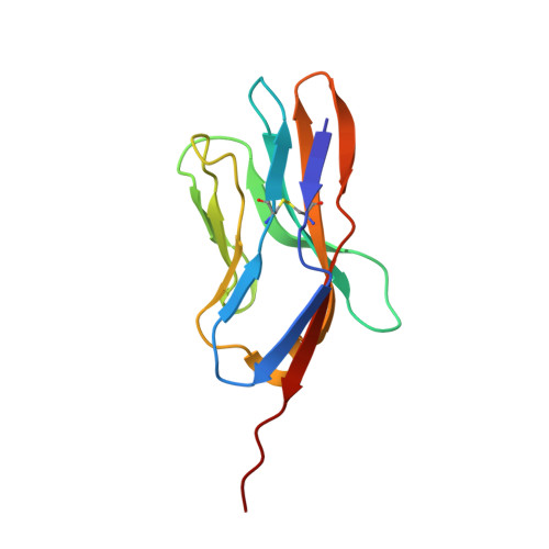
    The T-cell receptor (TCR) is a heterodimeric cell-surface protein consisting of two chains, alpha and beta, each of which is composed of a variable (V) and a constant (C) domain. Crystals of the isolated V(alpha) domain of the murine TCR 2C were grown by serendipity from a solution containing the extracellular domains of the intact TCR 2C and CD3 gamma epsilon-chains. The V(alpha) crystal structure shows how crystal packing can substitute for another V(alpha) domain in a different fashion from that observed in V(alpha)/V(alpha) homodimer and V(alpha)/V(beta) heterodimer structures. Significant conformational changes occur in the CDR3 and beta(3)beta(4) loops that normally form part of the dimer interface. The monomeric V(alpha) domain provides the unique opportunity to study the effect of dimerization on the conformation of the unliganded complementarity-determining regions (CDR) of a TCR. This structure of an individual V(alpha) module has implications for stability and bioengineering of isolated antibody and immunoglobulin domains.


  • Organizational Affiliation

    Department of Molecular Biology and Skaggs Institute for Chemical Biology (BCC-206), The Scripps Research Institute, 10550 North Torrey Pines Road, La Jolla, CA 92037, USA.


Macromolecules
Find similar proteins by:  (by identity cutoff)  |  3D Structure
Entity ID: 1
MoleculeChains Sequence LengthOrganismDetailsImage
CYTOTOXIC TCELL VALPHA DOMAIN115Mus musculusMutation(s): 0 
UniProt
Find proteins for P01738 (Mus musculus)
Explore P01738 
Go to UniProtKB:  P01738
Entity Groups  
Sequence Clusters30% Identity50% Identity70% Identity90% Identity95% Identity100% Identity
UniProt GroupP01738
Sequence Annotations
Expand
  • Reference Sequence
Small Molecules
Ligands 1 Unique
IDChains Name / Formula / InChI Key2D Diagram3D Interactions
NAG
Query on NAG

Download Ideal Coordinates CCD File 
B [auth A]2-acetamido-2-deoxy-beta-D-glucopyranose
C8 H15 N O6
OVRNDRQMDRJTHS-FMDGEEDCSA-N
Experimental Data & Validation

Experimental Data

  • Method: X-RAY DIFFRACTION
  • Resolution: 2.50 Å
  • R-Value Free: 0.233 
  • R-Value Work: 0.203 
  • R-Value Observed: 0.206 
  • Space Group: I 41 2 2
Unit Cell:
Length ( Å )Angle ( ˚ )
a = 104.032α = 90
b = 104.032β = 90
c = 63.09γ = 90
Software Package:
Software NamePurpose
DENZOdata reduction
SCALEPACKdata scaling
AMoREphasing
REFMACrefinement

Structure Validation

View Full Validation Report


Ligand Structure Quality Assessment 


Entry History 

Deposition Data

Revision History  (Full details and data files)

  • Version 1.0: 2001-12-05
    Type: Initial release
  • Version 1.1: 2008-04-27
    Changes: Version format compliance
  • Version 1.2: 2011-07-13
    Changes: Advisory, Refinement description, Version format compliance
  • Version 1.3: 2020-07-29
    Type: Remediation
    Reason: Carbohydrate remediation
    Changes: Data collection, Derived calculations, Structure summary
  • Version 1.4: 2023-08-09
    Changes: Data collection, Database references, Refinement description, Structure summary