1I7F

CRYSTAL STRUCTURE OF THE HSP33 DOMAIN WITH CONSTITUTIVE CHAPERONE ACTIVITY


Experimental Data Snapshot

  • Method: X-RAY DIFFRACTION
  • Resolution: 2.70 Å
  • R-Value Free: 0.290 
  • R-Value Work: 0.222 

wwPDB Validation   3D Report Full Report


This is version 1.3 of the entry. See complete history


Literature

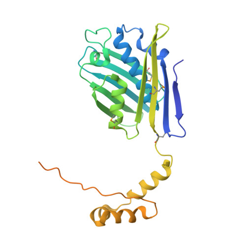
Crystal structure of proteolytic fragments of the redox-sensitive Hsp33 with constitutive chaperone activity

Kim, S.J.Jeong, D.G.Chi, S.W.Lee, J.S.Ryu, S.E.

(2001) Nat Struct Biol 8: 459-466

  • DOI: https://doi.org/10.1038/87639
  • Primary Citation of Related Structures:  
    1I7F

  • PubMed Abstract: 

    Heat shock protein 33 (Hsp33) inhibits aggregation of partially denatured proteins during oxidative stress. The chaperone activity of Hsp33 is unique among heat shock proteins because the activity is reversibly regulated by cellular redox status. We report here the crystal structure of the N-terminal region of Hsp33 fragments with constitutive chaperone activity. The structure reveals that the N-terminal portion of Hsp33 forms a tightly associated dimer formed by a domain crossover. A concave groove on the dimeric surface contains an elongated hydrophobic patch that could potentially bind denatured protein substrates. The termini of the subunits are located near the hydrophobic patch, indicating that the cleaved C-terminal domain may shield the hydrophobic patch in an inactive state. Two of the four conserved zinc-coordinating cysteines are in the end of the N-terminal domain, and the other two are in the cleaved C-terminal domain. The structural information and subsequent biochemical characterizations suggest that the redox switch of Hsp33 occurs by a reversible dissociation of the C-terminal regulatory domain through oxidation of zinc-coordinating cysteines and zinc release.


  • Organizational Affiliation

    Center for Cellular Switch Protein Structure, Korea Research Institute of Bioscience and Biotechnology, P. O. Box 115, Yusong, Taejon 305-600, South Korea.


Macromolecules
Find similar proteins by:  (by identity cutoff)  |  3D Structure
Entity ID: 1
MoleculeChains Sequence LengthOrganismDetailsImage
HEAT SHOCK PROTEIN 33292Escherichia coliMutation(s): 9 
Gene Names: HSP33
UniProt
Find proteins for P0A6Y5 (Escherichia coli (strain K12))
Explore P0A6Y5 
Go to UniProtKB:  P0A6Y5
Entity Groups  
Sequence Clusters30% Identity50% Identity70% Identity90% Identity95% Identity100% Identity
UniProt GroupP0A6Y5
Sequence Annotations
Expand
  • Reference Sequence
Small Molecules
Modified Residues  1 Unique
IDChains TypeFormula2D DiagramParent
MSE
Query on MSE
A
L-PEPTIDE LINKINGC5 H11 N O2 SeMET
Experimental Data & Validation

Experimental Data

  • Method: X-RAY DIFFRACTION
  • Resolution: 2.70 Å
  • R-Value Free: 0.290 
  • R-Value Work: 0.222 
  • Space Group: P 65 2 2
Unit Cell:
Length ( Å )Angle ( ˚ )
a = 77.293α = 90
b = 77.293β = 90
c = 193.893γ = 120
Software Package:
Software NamePurpose
SOLVEphasing
CNSrefinement
DENZOdata reduction
SCALEPACKdata scaling

Structure Validation

View Full Validation Report


Entry History 

Deposition Data

Revision History  (Full details and data files)

  • Version 1.0: 2001-05-09
    Type: Initial release
  • Version 1.1: 2008-04-27
    Changes: Version format compliance
  • Version 1.2: 2011-07-13
    Changes: Derived calculations, Version format compliance
  • Version 1.3: 2021-10-27
    Changes: Database references, Derived calculations