1I5R

TYPE 1 17-BETA HYDROXYSTEROID DEHYDROGENASE EM1745 COMPLEX


Experimental Data Snapshot

  • Method: X-RAY DIFFRACTION
  • Resolution: 1.60 Å
  • R-Value Free: 0.205 
  • R-Value Work: 0.185 

wwPDB Validation   3D Report Full Report


Ligand Structure Quality Assessment 


This is version 1.3 of the entry. See complete history


Literature

A concerted, rational design of type 1 17beta-hydroxysteroid dehydrogenase inhibitors: estradiol-adenosine hybrids with high affinity

Qiu, W.Campbell, R.L.Gangloff, A.Dupuis, P.Boivin, R.P.Tremblay, M.R.Poirier, D.Lin, S.X.

(2002) FASEB J 16: 1829-1831

  • DOI: https://doi.org/10.1096/fj.02-0026fje
  • Primary Citation of Related Structures:  
    1I5R

  • PubMed Abstract: 

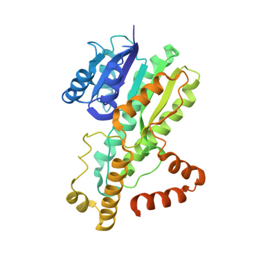
    Human estrogenic 17beta-hydroxysteroid dehydrogenase (17beta-HSD type 1) catalyzes the final step in the synthesis of active estrogens that stimulate the proliferation of breast cancer cells. Based on the initial premise to make use of the binding energies of both the substrate and cofactor sites, and molecular modeling starting from the enzyme structure, several estradiol-adenosine hybrids were designed and synthesized. Among these hybrids, EM-1745 with a linker of 8-CH2 groups is proved to be the best competitive inhibitor with a Ki of 3.0 +/- 0.8 nM. The crystal structure of the EM-1745 enzyme complex at 1.6 A provides evidence at atomic resolution of strong interactions between both the steroid and cofactor moieties and the enzyme molecule, as illustrated by a deltaA-weighted 2Fo-Fc electron density map contoured at 3.0 delta. The substrate entry loop is further stabilized in this complex compared with previous complexes of the enzyme. These results confirm our initial strategy of combining studies of structural biology and enzyme mechanism in the inhibitor design, which may be applied to other steroidogenic enzymes involved in human diseases.


  • Organizational Affiliation

    Oncology and Molecular Endocrinology Research Center, Laval University Medical Center (CHUL) and Laval University, Quebec, G1V 4G2, Canada.


Macromolecules
Find similar proteins by:  (by identity cutoff)  |  3D Structure
Entity ID: 1
MoleculeChains Sequence LengthOrganismDetailsImage
TYPE 1 17 BETA-HYDROXYSTEROID DEHYDROGENASE327Homo sapiensMutation(s): 0 
EC: 1.1.1.62
UniProt & NIH Common Fund Data Resources
Find proteins for P14061 (Homo sapiens)
Explore P14061 
Go to UniProtKB:  P14061
PHAROS:  P14061
GTEx:  ENSG00000108786 
Entity Groups  
Sequence Clusters30% Identity50% Identity70% Identity90% Identity95% Identity100% Identity
UniProt GroupP14061
Sequence Annotations
Expand
  • Reference Sequence
Small Molecules
Binding Affinity Annotations 
IDSourceBinding Affinity
HYC Binding MOAD:  1I5R Ki: 3 (nM) from 1 assay(s)
PDBBind:  1I5R Ki: 3 (nM) from 1 assay(s)
BindingDB:  1I5R Ki: 3 (nM) from 1 assay(s)
IC50: min: 8, max: 52 (nM) from 2 assay(s)
Experimental Data & Validation

Experimental Data

  • Method: X-RAY DIFFRACTION
  • Resolution: 1.60 Å
  • R-Value Free: 0.205 
  • R-Value Work: 0.185 
  • Space Group: C 1 2 1
Unit Cell:
Length ( Å )Angle ( ˚ )
a = 119.272α = 90
b = 42.706β = 99.16
c = 58.732γ = 90
Software Package:
Software NamePurpose
DENZOdata reduction
SCALEPACKdata scaling
REFMACrefinement

Structure Validation

View Full Validation Report


Ligand Structure Quality Assessment 


Entry History 

Deposition Data

Revision History  (Full details and data files)

  • Version 1.0: 2003-03-11
    Type: Initial release
  • Version 1.1: 2008-04-27
    Changes: Version format compliance
  • Version 1.2: 2011-07-13
    Changes: Derived calculations, Version format compliance
  • Version 1.3: 2023-08-09
    Changes: Data collection, Database references, Derived calculations, Refinement description