1I4M

Crystal structure of the human prion protein reveals a mechanism for oligomerization


Experimental Data Snapshot

  • Method: X-RAY DIFFRACTION
  • Resolution: 2.00 Å
  • R-Value Free: 0.253 
  • R-Value Work: 0.206 
  • R-Value Observed: 0.206 

wwPDB Validation   3D Report Full Report


This is version 1.2 of the entry. See complete history


Literature

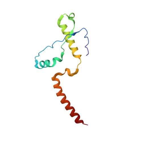
Crystal structure of the human prion protein reveals a mechanism for oligomerization.

Knaus, K.J.Morillas, M.Swietnicki, W.Malone, M.Surewicz, W.K.Yee, V.C.

(2001) Nat Struct Biol 8: 770-774

  • DOI: https://doi.org/10.1038/nsb0901-770
  • Primary Citation of Related Structures:  
    1I4M

  • PubMed Abstract: 

    The pathogenesis of transmissible encephalopathies is associated with the conversion of the cellular prion protein, PrP(C), into a conformationally altered oligomeric form, PrP(Sc). Here we report the crystal structure of the human prion protein in dimer form at 2 A resolution. The dimer results from the three-dimensional swapping of the C-terminal helix 3 and rearrangement of the disulfide bond. An interchain two-stranded antiparallel beta-sheet is formed at the dimer interface by residues that are located in helix 2 in the monomeric NMR structures. Familial prion disease mutations map to the regions directly involved in helix swapping. This crystal structure suggests that oligomerization through 3D domain-swapping may constitute an important step on the pathway of the PrP(C) --> PrP(Sc) conversion.


  • Organizational Affiliation

    Department of Molecular Cardiology and Center for Structural Biology, Lerner Research Institute, Cleveland Clinic Foundation, 9500 Euclid Avenue NB20, Cleveland, Ohio 44195, USA.


Macromolecules
Find similar proteins by:  (by identity cutoff)  |  3D Structure
Entity ID: 1
MoleculeChains Sequence LengthOrganismDetailsImage
MAJOR PRION PROTEIN108Homo sapiensMutation(s): 0 
UniProt & NIH Common Fund Data Resources
Find proteins for P04156 (Homo sapiens)
Explore P04156 
Go to UniProtKB:  P04156
PHAROS:  P04156
GTEx:  ENSG00000171867 
Entity Groups  
Sequence Clusters30% Identity50% Identity70% Identity90% Identity95% Identity100% Identity
UniProt GroupP04156
Sequence Annotations
Expand
  • Reference Sequence
Experimental Data & Validation

Experimental Data

  • Method: X-RAY DIFFRACTION
  • Resolution: 2.00 Å
  • R-Value Free: 0.253 
  • R-Value Work: 0.206 
  • R-Value Observed: 0.206 
  • Space Group: C 2 2 21
Unit Cell:
Length ( Å )Angle ( ˚ )
a = 85.441α = 90
b = 85.707β = 90
c = 40.501γ = 90
Software Package:
Software NamePurpose
MLPHAREphasing
CNSrefinement
DENZOdata reduction
SCALEPACKdata scaling

Structure Validation

View Full Validation Report


Entry History 

Deposition Data

Revision History  (Full details and data files)

  • Version 1.0: 2002-02-27
    Type: Initial release
  • Version 1.1: 2008-04-27
    Changes: Version format compliance
  • Version 1.2: 2011-07-13
    Changes: Derived calculations, Version format compliance