1HVH

NONPEPTIDE CYCLIC CYANOGUANIDINES AS HIV PROTEASE INHIBITORS


Experimental Data Snapshot

  • Method: X-RAY DIFFRACTION
  • Resolution: 1.80 Å
  • R-Value Work: 0.188 
  • R-Value Observed: 0.188 

wwPDB Validation   3D Report Full Report


This is version 1.4 of the entry. See complete history


Literature

Nonpeptide cyclic cyanoguanidines as HIV-1 protease inhibitors: synthesis, structure-activity relationships, and X-ray crystal structure studies.

Jadhav, P.K.Woerner, F.J.Lam, P.Y.Hodge, C.N.Eyermann, C.J.Man, H.W.Daneker, W.F.Bacheler, L.T.Rayner, M.M.Meek, J.L.Erickson-Viitanen, S.Jackson, D.A.Calabrese, J.C.Schadt, M.Chang, C.H.

(1998) J Med Chem 41: 1446-1455

  • DOI: https://doi.org/10.1021/jm970524i
  • Primary Citation of Related Structures:  
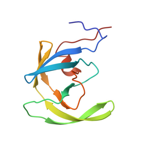
    1HVH

  • PubMed Abstract: 

    Comparison of the high-resolution X-ray structures of the native HIV-1 protease and its complexes with the inhibitors suggested that the enzyme flaps are flexible. The movement at the tip of the flaps could be as large as 7 A. On the basis of this observation, cyclic cyanoguanidines have been designed, synthesized, and evaluated as HIV-1 protease (PR) inhibitors. Cyclic cyanoguanidines were found to be very potent inhibitors of HIV-1 protease. The choice of cyclic cyanoguanidines over cyclic guanidines was based on the reduced basicity of the former. X-ray structure studies of the HIV PR complex with cyclic cyanoguanidine demonstrated that in analogy to cyclic urea, cyclic cyanoguanidines also displace the unique structural water molecule. The structure-activity relationship of the cyclic cyanoguanidines is compared with that of the corresponding cyclic urea analogues. The differences in binding constants of the two series of compounds have been rationalized using high-resolution X-ray structure information.


  • Organizational Affiliation

    Chemical and Physical Sciences Department, The DuPont Merck Pharmaceutical Company, Experimental Station, P.O. Box 80500, Wilmington, Delaware 19880-0500, USA. Prabhakar.K.Jadhav@dupontmerck.com


Macromolecules
Find similar proteins by:  (by identity cutoff)  |  3D Structure
Entity ID: 1
MoleculeChains Sequence LengthOrganismDetailsImage
HIV-1 PROTEASE
A, B
99Human immunodeficiency virus 1Mutation(s): 0 
EC: 3.4.23.16
UniProt
Find proteins for P04585 (Human immunodeficiency virus type 1 group M subtype B (isolate HXB2))
Explore P04585 
Go to UniProtKB:  P04585
Entity Groups  
Sequence Clusters30% Identity50% Identity70% Identity90% Identity95% Identity100% Identity
UniProt GroupP04585
Sequence Annotations
Expand
  • Reference Sequence
Small Molecules
Ligands 1 Unique
IDChains Name / Formula / InChI Key2D Diagram3D Interactions
Q82
Query on Q82

Download Ideal Coordinates CCD File 
C [auth B]{[4-R(-4-ALPHA,5-ALPHA,6-BETA,7-BETA)]-HEXAHYDRO-5,6-BIS(HYDROXY)-1,3-BIS(4-HYDROXYMETHYL)METHYL]-4,7-BIS(PHENYLMETHYL) -2H-1,3-DIAZEPIN-2-YLIDENE]CYANAMIDE}
C36 H38 N4 O4
VLCZFBGXHAKRLP-WDKGQIBQSA-N
Binding Affinity Annotations 
IDSourceBinding Affinity
Q82 Binding MOAD:  1HVH Ki: 11 (nM) from 1 assay(s)
BindingDB:  1HVH Ki: 11 (nM) from 1 assay(s)
PDBBind:  1HVH Ki: 11 (nM) from 1 assay(s)
Experimental Data & Validation

Experimental Data

  • Method: X-RAY DIFFRACTION
  • Resolution: 1.80 Å
  • R-Value Work: 0.188 
  • R-Value Observed: 0.188 
  • Space Group: P 61
Unit Cell:
Length ( Å )Angle ( ˚ )
a = 63.22α = 90
b = 63.22β = 90
c = 83.68γ = 120
Software Package:
Software NamePurpose
X-PLORmodel building
X-PLORrefinement
X-PLORphasing

Structure Validation

View Full Validation Report


Entry History 

Deposition Data

  • Released Date: 1998-12-30 
  • Deposition Author(s): Chang, C.-H.

Revision History  (Full details and data files)

  • Version 1.0: 1998-12-30
    Type: Initial release
  • Version 1.1: 2008-03-24
    Changes: Version format compliance
  • Version 1.2: 2011-07-13
    Changes: Version format compliance
  • Version 1.3: 2017-11-29
    Changes: Derived calculations, Other
  • Version 1.4: 2023-08-09
    Changes: Database references, Derived calculations, Refinement description, Structure summary