1HQ1

STRUCTURAL AND ENERGETIC ANALYSIS OF RNA RECOGNITION BY A UNIVERSALLY CONSERVED PROTEIN FROM THE SIGNAL RECOGNITION PARTICLE


Experimental Data Snapshot

  • Method: X-RAY DIFFRACTION
  • Resolution: 1.52 Å
  • R-Value Free: 0.199 
  • R-Value Work: 0.151 
  • R-Value Observed: 0.151 

wwPDB Validation   3D Report Full Report


This is version 1.4 of the entry. See complete history


Literature

Structural and energetic analysis of RNA recognition by a universally conserved protein from the signal recognition particle.

Batey, R.T.Sagar, M.B.Doudna, J.A.

(2001) J Mol Biol 307: 229-246

  • DOI: https://doi.org/10.1006/jmbi.2000.4454
  • Primary Citation of Related Structures:  
    1HQ1

  • PubMed Abstract: 

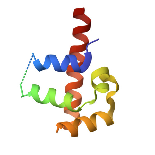
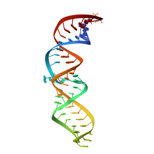
    The signal recognition particle (SRP) is a ribonucleoprotein complex responsible for targeting proteins to the endoplasmic reticulum in eukarya or to the inner membrane in prokarya. The crystal structure of the universally conserved RNA-protein core of the Escherichia coli SRP, refined here to 1.5 A resolution, revealed minor groove recognition of the 4.5 S RNA component by the M domain of the Ffh protein. Within the RNA, nucleotides comprising two phylogenetically conserved internal loops create a unique surface for protein recognition. To determine the energetic importance of conserved nucleotides for SRP assembly, we measured the affinity of the M domain for a series of RNA mutants. This analysis reveals how conserved nucleotides within the two internal loop motifs establish the architecture of the macromolecular interface and position essential functional groups for direct recognition by the protein.


  • Organizational Affiliation

    Department of Molecular Biophysics and Biochemistry and Howard Hughes Medical Institute, Yale University, P.O. Box 208114, New Haven, CT 06520-8814, USA.


Macromolecules

Find similar proteins by:  (by identity cutoff)  |  3D Structure
Entity ID: 2
MoleculeChains Sequence LengthOrganismDetailsImage
SIGNAL RECOGNITION PARTICLE PROTEINB [auth A]105Escherichia coliMutation(s): 1 
UniProt
Find proteins for P0AGD7 (Escherichia coli (strain K12))
Explore P0AGD7 
Go to UniProtKB:  P0AGD7
Entity Groups  
Sequence Clusters30% Identity50% Identity70% Identity90% Identity95% Identity100% Identity
UniProt GroupP0AGD7
Sequence Annotations
Expand
  • Reference Sequence
Find similar nucleic acids by:  (by identity cutoff)  |  3D Structure
Entity ID: 1
MoleculeChains LengthOrganismImage
4.5S RNA DOMAIN IVA [auth B]49N/A
Sequence Annotations
Expand
  • Reference Sequence
Experimental Data & Validation

Experimental Data

  • Method: X-RAY DIFFRACTION
  • Resolution: 1.52 Å
  • R-Value Free: 0.199 
  • R-Value Work: 0.151 
  • R-Value Observed: 0.151 
  • Space Group: C 1 2 1
Unit Cell:
Length ( Å )Angle ( ˚ )
a = 136.555α = 90
b = 78.337β = 96.24
c = 32.872γ = 90
Software Package:
Software NamePurpose
AMoREphasing
SHELXL-97refinement
DENZOdata reduction
SCALEPACKdata scaling

Structure Validation

View Full Validation Report


Entry History 

Deposition Data

Revision History  (Full details and data files)

  • Version 1.0: 2001-01-03
    Type: Initial release
  • Version 1.1: 2008-04-27
    Changes: Version format compliance
  • Version 1.2: 2011-07-13
    Changes: Version format compliance
  • Version 1.3: 2021-10-27
    Changes: Database references, Derived calculations
  • Version 1.4: 2023-08-09
    Changes: Data collection, Refinement description