1HBZ

Catalase from Micrococcus lysodeikticu


Experimental Data Snapshot

  • Method: X-RAY DIFFRACTION
  • Resolution: 1.50 Å
  • R-Value Free: 0.120 
  • R-Value Work: 0.092 

wwPDB Validation   3D Report Full Report


Ligand Structure Quality Assessment 


This is version 1.3 of the entry. See complete history


Literature

Three-Dimensional Structure of Catalase from Micrococcus Lysodeikticus at 1.5A Resolution

Murshudov, G.N.Melik-Adamyan, W.R.Grebenko, A.I.Barynin, V.V.Vagin, A.A.Vainshtein, B.K.Dauter, Z.Wilson, K.S.

(1992) FEBS Lett 312: 127

  • DOI: https://doi.org/10.1016/0014-5793(92)80919-8
  • Primary Citation of Related Structures:  
    1HBZ

  • PubMed Abstract: 

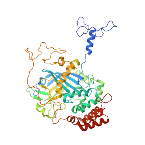
    The three-dimensional crystal structure of catalase from Micrococcus lysodeikticus has been solved by multiple isomorphous replacement and refined at 1.5 A resolution. The subunit of the tetrameric molecule of 222 symmetry consists of a single polypeptide chain of about 500 amino acid residues and one haem group. The crystals belong to space group P4(2)2(1)2 with unit cell parameters a = b = 106.7 A, c = 106.3 A, and there is one subunit of the tetramer per asymmetric unit. The amino acid sequence has been tentatively determined by computer graphics model building and comparison with the known three-dimensional structure of beef liver catalase and sequences of several other catalases. The atomic model has been refined by Hendrickson and Konnert's least-squares minimisation against 94,315 reflections between 8 A and 1.5 A. The final model consists of 3,977 non-hydrogen atoms of the protein and haem group, 426 water molecules and one sulphate ion. The secondary and tertiary structures of the bacterial catalase have been analyzed and a comparison with the structure of beef liver catalase has been made.


  • Organizational Affiliation

    Institute of Crystallography, Russian Academy of Sciences, Moscow.


Macromolecules
Find similar proteins by:  (by identity cutoff)  |  3D Structure
Entity ID: 1
MoleculeChains Sequence LengthOrganismDetailsImage
CATALASE498Micrococcus luteusMutation(s): 0 
EC: 1.11.1.6
UniProt
Find proteins for P29422 (Micrococcus luteus)
Explore P29422 
Go to UniProtKB:  P29422
Entity Groups  
Sequence Clusters30% Identity50% Identity70% Identity90% Identity95% Identity100% Identity
UniProt GroupP29422
Sequence Annotations
Expand
  • Reference Sequence
Small Molecules
Ligands 2 Unique
IDChains Name / Formula / InChI Key2D Diagram3D Interactions
HEM
Query on HEM

Download Ideal Coordinates CCD File 
B [auth A]PROTOPORPHYRIN IX CONTAINING FE
C34 H32 Fe N4 O4
KABFMIBPWCXCRK-RGGAHWMASA-L
SO4
Query on SO4

Download Ideal Coordinates CCD File 
C [auth A]SULFATE ION
O4 S
QAOWNCQODCNURD-UHFFFAOYSA-L
Experimental Data & Validation

Experimental Data

  • Method: X-RAY DIFFRACTION
  • Resolution: 1.50 Å
  • R-Value Free: 0.120 
  • R-Value Work: 0.092 
  • Space Group: P 42 21 2
Unit Cell:
Length ( Å )Angle ( ˚ )
a = 106.7α = 90
b = 106.7β = 90
c = 106.25γ = 90
Software Package:
Software NamePurpose
REFMACrefinement
MOSFLMdata scaling

Structure Validation

View Full Validation Report


Ligand Structure Quality Assessment 


Entry History 

Deposition Data

Revision History  (Full details and data files)

  • Version 1.0: 2001-05-24
    Type: Initial release
  • Version 1.1: 2011-05-07
    Changes: Version format compliance
  • Version 1.2: 2011-07-13
    Changes: Version format compliance
  • Version 1.3: 2019-07-24
    Changes: Data collection