1HAU

X-RAY STRUCTURE OF A BLUE COPPER NITRITE REDUCTASE AT HIGH PH AND IN COPPER FREE FORM AT 1.9 A RESOLUTION


Experimental Data Snapshot

  • Method: X-RAY DIFFRACTION
  • Resolution: 1.90 Å
  • R-Value Free: 0.190 
  • R-Value Work: 0.154 
  • R-Value Observed: 0.156 

wwPDB Validation   3D Report Full Report


This is version 1.2 of the entry. See complete history


Literature

X-Ray Structure of a Blue Copper Nitrite Reductase at High Ph and in Copper-Free Form at 1.9 A Resolution

Ellis, M.J.Dodd, F.E.Strange, R.W.Prudencio, M.Sawers, G.Eady, R.R.Hasnain, S.S.

(2001) Acta Crystallogr D Biol Crystallogr 57: 1110

  • DOI: https://doi.org/10.1107/s0907444901008654
  • Primary Citation of Related Structures:  
    1HAU, 1HAW

  • PubMed Abstract: 

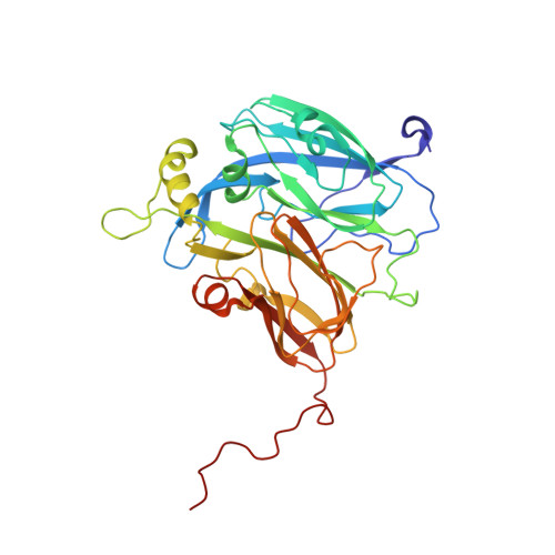
    Copper-containing nitrite reductases possess a trimeric structure where the catalytic Cu site, located at the monomer-monomer interface, resembles the catalytic sites of a number of Zn enzymes. Nitrite reductase from Alcaligenes xylosoxidans has optimum activity at pH 5.2 which decreases to a negligible level at pH 8. The structure of this nitrite reductase has previously been determined at pH 4.6. It has now been crystallized under new conditions at pH 8.5. Its crystallographic structure provides a structural explanation for the greatly reduced activity of the enzyme at high pH. Characterization of overexpressed protein in solution by EXAFS suggested that the protein lacked Cu in the catalytic type 2 Cu site and that the site was most probably occupied by Zn. Using the anomalous signals from Cu and Zn, the crystal structure revealed that the expressed protein was devoid of Cu in the catalytic site and that only a trace amount (<10%) of Zn was present at this site in the crystal. Despite the close structural similarity of the catalytic site to a number of Zn enzymes, these data suggest that Zn, if it binds at the catalytic copper site, binds weakly in nitrite reductase.


  • Organizational Affiliation

    Synchrotron Radiation Department, CLRC Daresbury Laboratory, Warrington WA4 4AD, England.


Macromolecules
Find similar proteins by:  (by identity cutoff)  |  3D Structure
Entity ID: 1
MoleculeChains Sequence LengthOrganismDetailsImage
DISSIMILATORY COPPER-CONTAINING NITRITE REDUCTASE336Achromobacter xylosoxidansMutation(s): 0 
EC: 1.7.99.3 (PDB Primary Data), 1.7.2.1 (PDB Primary Data)
UniProt
Find proteins for O68601 (Alcaligenes xylosoxydans xylosoxydans)
Explore O68601 
Go to UniProtKB:  O68601
Entity Groups  
Sequence Clusters30% Identity50% Identity70% Identity90% Identity95% Identity100% Identity
UniProt GroupO68601
Sequence Annotations
Expand
  • Reference Sequence
Small Molecules
Ligands 1 Unique
IDChains Name / Formula / InChI Key2D Diagram3D Interactions
CU
Query on CU

Download Ideal Coordinates CCD File 
B [auth A],
C [auth A]
COPPER (II) ION
Cu
JPVYNHNXODAKFH-UHFFFAOYSA-N
Experimental Data & Validation

Experimental Data

  • Method: X-RAY DIFFRACTION
  • Resolution: 1.90 Å
  • R-Value Free: 0.190 
  • R-Value Work: 0.154 
  • R-Value Observed: 0.156 
  • Space Group: P 63
Unit Cell:
Length ( Å )Angle ( ˚ )
a = 80.978α = 90
b = 80.978β = 90
c = 99.781γ = 120
Software Package:
Software NamePurpose
REFMACrefinement
DENZOdata reduction
CCP4data scaling
AMoREphasing

Structure Validation

View Full Validation Report


Entry History 

Deposition Data

Revision History  (Full details and data files)

  • Version 1.0: 2001-08-02
    Type: Initial release
  • Version 1.1: 2012-05-09
    Changes: Atomic model, Data collection, Database references, Derived calculations, Non-polymer description, Other, Structure summary, Version format compliance
  • Version 1.2: 2023-12-13
    Changes: Data collection, Database references, Derived calculations, Other, Refinement description